Background: While preclinical and early clinical studies suggest the safety and efficacy of hepatic denervation (HDN) and larger clinical trials are underway, detailed anatomical understanding of hepatic perivascular nerves remains scarce.
Aims: To characterize periarterial nerve fibers along the hepatic artery and identify anatomically favorable target regions for catheter-based HDN.
Methods: Hepatic arteries with surrounding tissue from the origin at the coeliac trunk to the intrahepatic entry were excised from ten body donors and examined histologically. Immunofluorescence staining was performed to characterize sympathetic nerve fibers.
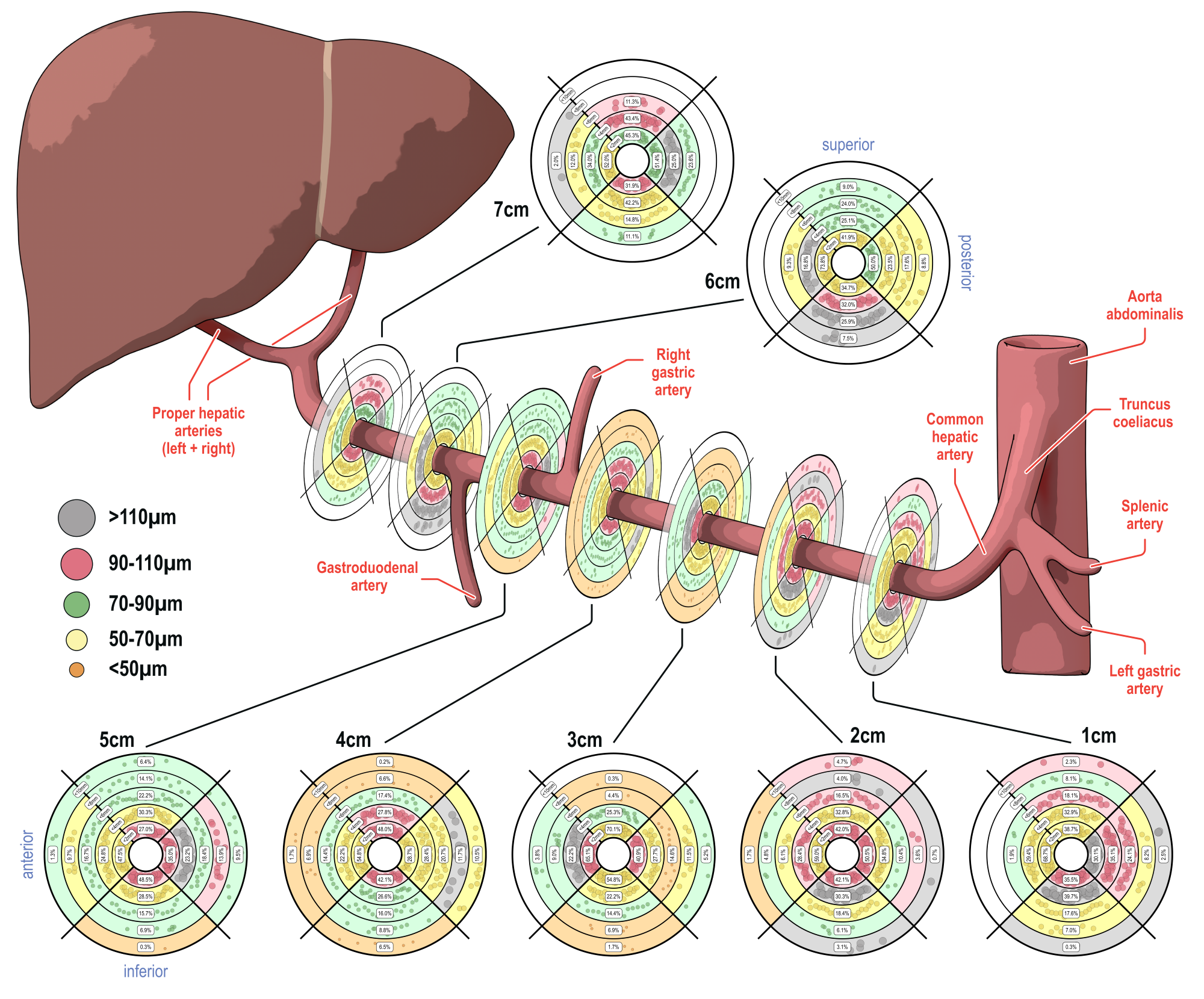
Results: A total of 12,841 nerves were analyzed. Median nerve fiber diameter decreased progressively from 93 µm [IQR 54–165] at 1 cm to 72 µm [IQR 48–120] at 7 cm distal to the coeliac trunk (p<0.001). The largest nerve fibers were located in the posterior and inferior quadrants, each measuring 85 µm [IQR 48–176 and 48–158]. Mean nerve fiber density was significantly higher within the proximal 1–3 cm compared to distal segments 4–7 cm from the coeliac trunk (233±124 vs. 159±76 nerves/cm², p=0.003). Lumen-nerve distance was significantly smaller within the proximal 3 cm compared with distal segments 4–7 cm from the coeliac trunk (2.03 mm [IQR 1.23–3.58] vs. 2.57 mm [IQR 1.26–4.67], p<0.001). Compared with other quadrants, the shortest lumen–nerve distance was observed in the anterior quadrant (1.75 mm [IQR 1.10–3.22], p<0.001).
Conclusions: The hepatic artery is densely innervated with a favorable distribution pattern in the proximal region characterized by larger nerve fibers, higher density, and shorter lumen–nerve distances. These findings may guide the safe and effective application of HDN in humans.