Background: Non-invasive imaging derived myocardial blood flow (MBF) quantification can be applied to identify coronary artery disease. Recently, fully automated MBF assessment by cardiac MRI has become available. There are not only limited but also controversial data regarding the impact of cardiovascular risk factors and patient´s characteristics (age, BMI, and sex) on MBF at rest and during hyperemia.
Objectives: The aim of this study was to investigate the impact of cardiovascular risk factors on MBF in individuals without regional inducible ischemia suggesting absence of obstructive (epicardial) coronary disease.
Methods: Consecutive patients who underwent stress CMR at our institution in 2023 were included in this study. First pass perfusion was assessed visually as well as quantitatively using an artificial intelligence based fully automated inline MRI-sequence. All patients included had a normal S-CMR exam by routine qualitative assessment (i.e. absence of: LGE, perfusion defects, valvular and structural heart disease). Only patients with sufficient vasodilator stress (defined as: stress MBF (SMBF) >1.43ml/g/min in at least 1 AHA segment, heart rate increase >10bpm or blood pressure drop >10 mmHg) were included. Rest MBF (RMBF) was normalised to rate pressure product in order to minimise impact of hemodynamic differences during rest condition.
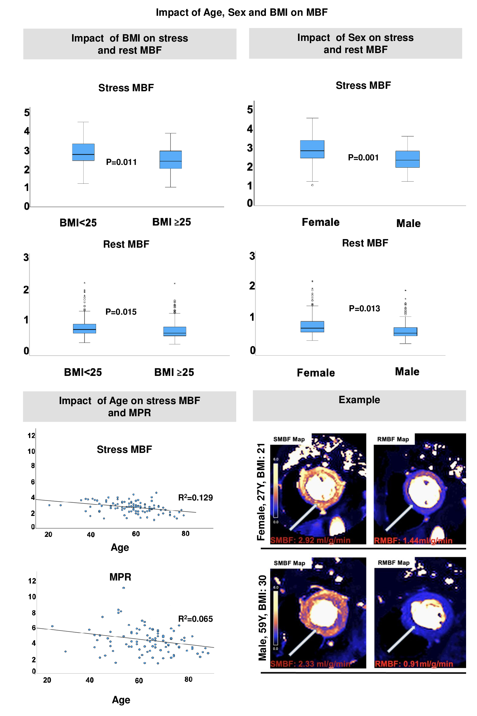
Results: Four hundred thirty-one individuals (age 62(55-68) years, 48.7% male) were included in this registry analysis. Ninety-eight patients (age 59(51.7-75.6) years, 40.8% male) had no known cardiovascular risk factors. The presence of cardiovascular risk factors was significantly associated with abnormal SMBF (<2.25ml/g/min.), especially arterial hypertension (RR: 0.704 (0.504-0.918), p=0.008), diabetes (RR: 0.631(0.46-0.86), p= 0.012) and hyperlipidemia (RR: 0.759 (0.59-0.97), p=0.032). Moreover, in a sub-analysis of individuals without risk factors, age, BMI, and sex were associated with differences in MBF. SMBF and RMBF were significantly lower in obese patients (SMBF: 2.789 (2.457-3.350) vs. 2.429 (1.995-2.993) ml/g/min, p=0.011; RMBF: 0.664 (0.566-0.853) vs. 0.548 (0.486-0.782) ml/g/min, p=0.015). Women demonstrated higher MBF values compared to men (SMBF 2.82 (2.431-3.377) vs. 2.328 (1.923-2.801) ml/g/min, p=0.001; RMBF 0.662 (0.559-0.860) vs. 0.544 (0.487-0.746) ml/g/min, p=0.013). Finally, we observed a linear decline of SMBF and myocardial perfusion reserve (MPR) with increasing age (SMBF: R2 =0.129, F=14.243, p<0.001, b0=3.996, b1=-0.023).
Conclusions : Automated MBF quantification by CMR demonstrates significant impact of cardiovascular risk factors and patient characteristics on myocardial perfusion in patients with otherwise normal CMR exam. These results support an important role of coronary microvascular disease in the absence of epicardial stenosis or structural heart disease and highlights the complexity determining universal MBF cut-off values.