Background
The P-Wave Area Index (PWAi) has been identified as a marker of atrial myopathy and low-voltage areas (LVA). This study aimed to investigate the association between PWAi and atrial fibrillation (AF) recurrence following pulmonary vein isolation (PVI).
Methods
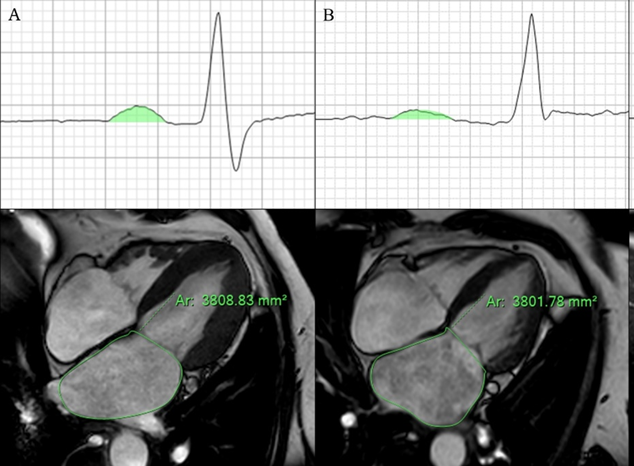
Patients undergoing first-time PVI were included in the analysis. A 12-lead electrocardiogram (ECG) in sinus rhythm was recorded prior to the procedure, and cardiac magnetic resonance imaging (CMR) (Ingenia 1.5T Philips) was performed to assess left atrial (LA) area. The PWAi was calculated by indexing the P-wave area in leads II to the LA area (PWAi = P-wave area / LA area, Figure 1). The primary outcome was AF recurrence during follow-up. Kaplan-Meier survival analysis was used to evaluate the association between PWAi and recurrence risk.
Results
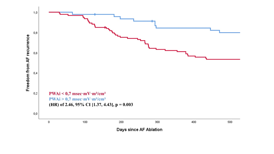
A total of 187 consecutive patients (mean age 65 years, 45% female, 51% persistent AF, 21% LVA) were included. During follow-up, 41% of patients experienced AF recurrences. Kaplan-Meier analysis demonstrated that a lower PWAi (<0,7 ms*mv*m²/cm²) was significantly associated with a higher recurrence rate (log-rank p 0.003), and was the most robust predictor among all analyzed parameters (OR: 2.333, p 0.034; Table 1), whereas those with higher PWAi values exhibited greater long-term rhythm stability following PVI (Figure 2).
Conclusion
The P-Wave Area Index is a predictor of AF recurrence after PVI. Its incorporation into preprocedural risk assessment may help to improve patient selection, individualize AF treatment strategies and may indicate a closer patient follow up.
Figure 1:
P Wave Area in lead II and left atrial Area in patients (a) without and (b) with present atrial myopathy and AF recurrence
|
Figure 2:
Kaplan-Meier survival analysis for P-Wave Area index in patients with AF recurrences
|
Table 1:
Multivariate Analysis for predefined categorial outcome variables
|
|
| |
Odds Ratio
|
95% Confidence Intervall
for Odds Ratio
|
p- value
|
|
|
|
lower
|
upper
|
|
|
female gender
|
1.651
|
0.799
|
3.415
|
0.176
|
|
|
persistent AF
|
1.871
|
0.924
|
3.786
|
0.082
|
|
|
GFR (ml/min)
|
0.982
|
0.960
|
1,005
|
0.128
|
|
|
LVA
|
1.099
|
0.416
|
2.905
|
0.849
|
|
|
PWAi
≤ 0.7 ms*mv*m²/cm²
|
2.333
|
1.068
|
5.096
|
0.034
|
|
|
|
|
|
|
|
|
|
|
| |
|
|
|
|
|
|
|
|
|
|
|
.
|


