Background: No cause is found in up to a third of patients with ischemic stroke, and one of the major possible occult causes are cardiac embolic sources. Left atrial appendage (LAA) is one of the main anatomical sources of cardiac embolism, which in many circumstances is not investigated in routine etiological stroke work-up.
Aim: The goal of this study was to analyse the diagnostic accuracy of cardiac photon-counting detector computed tomography (PCD-CT) integrated in the acute stroke imaging protocol for detection of LAA thrombus.
Methods: We conducted a retrospective single-centre diagnostic accuracy study of consecutive acute ischemic stroke or transient ischemic attack patients who underwent both emergent cardiac PCD-CT (
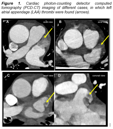
Figure 1) and transesophageal echocardiography (TEE) (
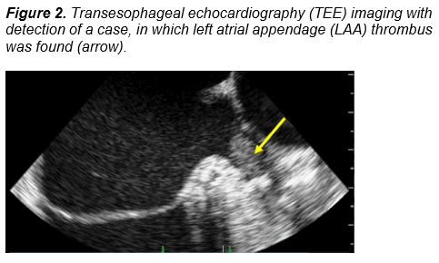
Figure 2) during index hospital admission. Cardiac PCD-CT and TEE images were independently assessed by two pairs of observers for detection of LAA thrombus. Patients with and without LAA thrombus as defined by PCD-CT were compared. Accuracy, sensitivity, specificity, positive and negative predictive value, and positive likelihood ratio for detection of LAA thrombus by cardiac PCD-CT were calculated (TEE used as gold-standard).
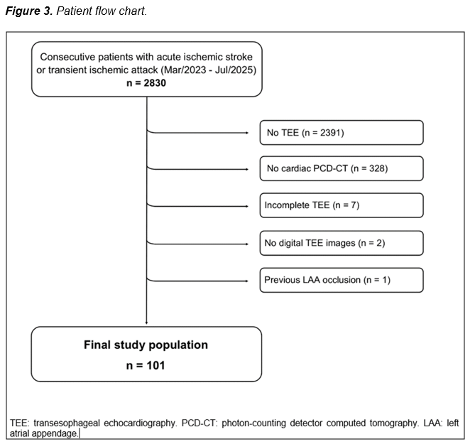
Results: We included 101 of 2830 screened patients, 42 were female (42%), median age was 59 years, of which 87 patients had acute ischemic stroke (86%) (
Figure 3). Median time between cardiac PCD-CT and TEE was 2 days. LAA thrombus defined by cardiac PCD-CT was present in 12 patients (12%). Patients with LAA thrombus were older (58 versus 74 years, p<0.001) and more frequently had atrial fibrillation (83.3% versus 23.6%, p<0.001) (
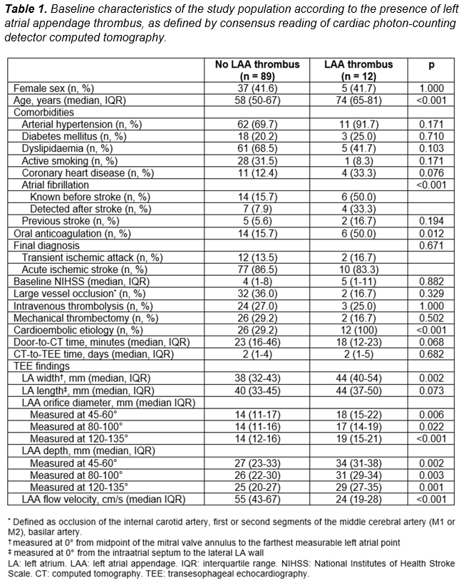
Table 1). Diagnostic accuracy of cardiac PCD-CT for detection of LAA thrombus was 96% (95% confidence interval [95%CI=90-99%), sensitivity was 90% (95%CI=56-100), specificity was 97% (95%CI=91-99%), positive predictive value was 75% (95%CI=49-90%), negative predictive value was 99% (95%CI=93-100%) and positive likelihood ratio was 27.3 (95%CI=8.8-84.7).
Conclusions: Cardiac PCD-CT integrated as part of the acute stroke imaging protocol has an excellent diagnostic accuracy for detecting LAA thrombi when compared with TEE as the gold standard. The findings of this study argue for the widespread implementation of cardiac CT in this setting for improving the etiological non-invasive investigation of stroke.