Background: Three-dimensional cardiac reconstruction platforms are increasingly used for pre-procedural planning in catheter ablation of ventricular tachycardia (VT). inHeart and CARDIO-RT both generate CT-derived myocardial wall thickness models, but whether they yield equivalent substrate identification remains unclear.
Methods: A single-centre observer-blinded study was performed using anonymised 3D cardiac models. Ten experienced electrophysiology operators independently identified one candidate VT isthmus per case (n=10) on models generated by each platform. Inter-platform agreement was quantified using the Jaccard similarity coefficient and Dice Similarity Coefficient (DSC), both calculated pairwise per operator, and the Weighted Overlap Coefficient (WOC), calculated at group level with operator-frequency weighting. Interobserver agreement within each platform was assessed using Fleiss' kappa.
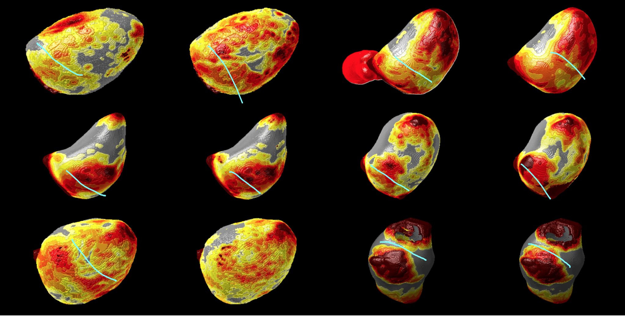
Figure 1. Representative 3D cardiac reconstructions (inHeart and CARDIO-RT). Cyan lines indicate operator-identified VT isthmus regions. Colour scale reflects CT-derived myocardial wall thickness (grey: normal/thick; yellow–red: thin/scar).
Results: WOC exceeded Jaccard in all cases (mean difference 0.143), indicating that majority-consensus operator selections were robustly reproduced across platforms. Case-level agreement ranged from complete concordance (Jaccard = 1.000) to substantial divergence (Jaccard = 0.300), reflecting differences in anatomical substrate complexity. Fleiss' kappa indicated fair interobserver agreement within both platforms, with no significant difference between systems.
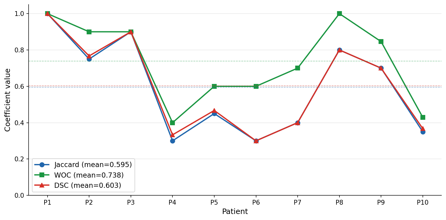
Figure 2. Inter-platform agreement by patient: Jaccard (blue), WOC (green), DSC (red). Dashed lines = respective means.
Conclusion: inHeart and CARDIO-RT demonstrated moderate-to-good inter-platform agreement in VT isthmus identification, with no systematic bias favouring either system. Residual discordance was attributable to anatomical substrate complexity rather than platform-specific differences. Both platforms appear clinically equivalent as visualisation tools for VT substrate assessment.



