Introduction
White matter hyperintensities (WMH), an MRI marker of small vessel disease, are increased in individuals with atrial fibrillation (AF) and associate with impaired neurocognitive function, dementia, and stroke. However, it remains unclear whether WMH are a consequence of AF or precede its clinical diagnosis. We therefore investigated if WMH manifest in individuals at increased risk of AF based on clinical, imaging, and genetic information, but without the diagnosis of AF.
Methods
This research was conducted using the UK Biobank Resource (Application No. 2964). We included 58.925 participants of the imaging cohort who underwent heart and brain MRI and had available information on WMH. We excluded those with a known diagnosis of AF or stroke at the time of imaging. Clinical risk for AF was quantified by the CHARGE-AF score, which weighs comorbidities and clinical factors to determine the 5-year risk for AF. Increased risk from imaging was defined as presence of atrial cardiomyopathy by three cardiac MRI measures: maximum left atrial volume index >60 ml/m2; minimum left atrial volume index >30 ml/m2; left atrial emptying fraction <45%. Genetic risk for AF was determined by an AF polygenic risk score. We constructed logistic regression models for the highest vs. the lower four quintiles of log-transformed WMH indexed to intracranial volume as the outcome and clinical, imaging, and genetic information as predictors.
Results
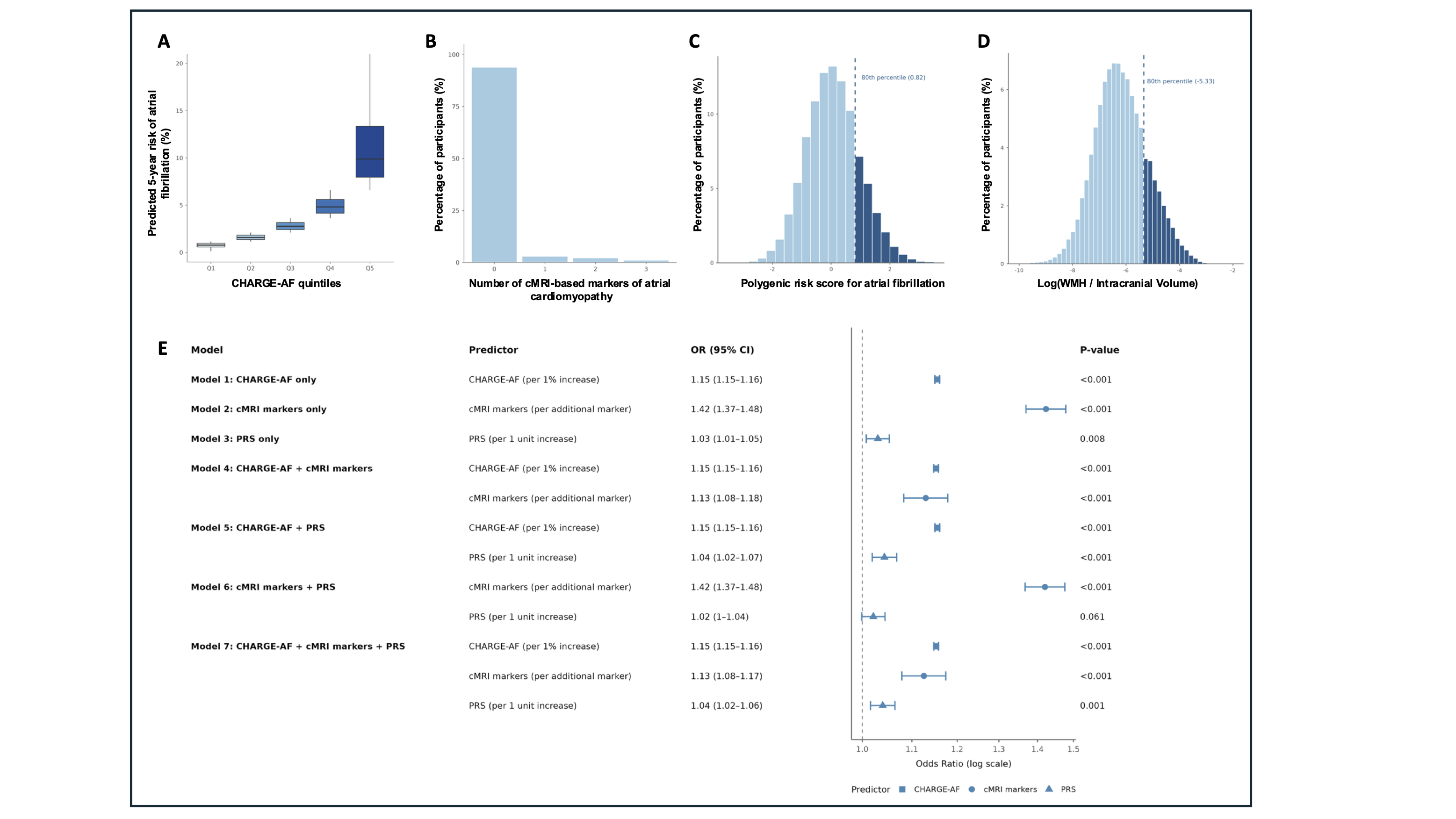
Participants had a mean age of 65.4±7.8 years, 54.2% were female, and 96.4% were of white ethnicity. The median CHARGE-AF score was 2.8% [1.4–5.6] and the median CHA2DS2-VASc Score was 2 [1-2]. Common cardiovascular risk factors included hypertension (29.4%), coronary artery disease (5.5%), and diabetes mellitus (5.1%). The distributions of the clinical, imaging, and genetic predictors, and the distribution of WMH are visualized in Figures 1A-1D. Clinical, imaging, and genetic predictors were significantly associated with a high WMH burden (Figure 1E). Per 1% increase of the CHARGE-AF score, the odds ratio (OR) was 1.15 (95%CI 1.15-1.16, p<0.001). Per additional imaging marker, the OR was 1.42 (95%CI 1.37-1.48, p<0.001). Per unit increase of the AF polygenic risk score, the OR was 1.03 (95%CI 1.01-1.05, p=0.008, Fig. 1E) A combined model yielded similar results. The area under the ROC curve to predict a high WMH burden was 0.75.
Conclusion
In a large, well-characterized population-based cohort, clinical, imaging, and genetic risk of AF predicted an excess of WMH in individuals without known AF. The excess in WMH burden may reflect clinically undiagnosed AF or may indicate the consequences of an advanced atrial cardiomyopathy. The interplay between AF risk, AF, and WMH should be subject to further study.