Background
Epsilon waves are considered a hallmark of arrhythmogenic right ventricular cardiomyopathy (ARVC), but they are not disease-specific and may occur in other conditions affecting the right ventricle, thereby mimicking ARVC. We report a diagnostically challenging case of patient with ventricular tachycardia (VT) and prominent epsilon waves in sinus rhythm, ultimately revealing an underlying systemic disease.
Case Summary
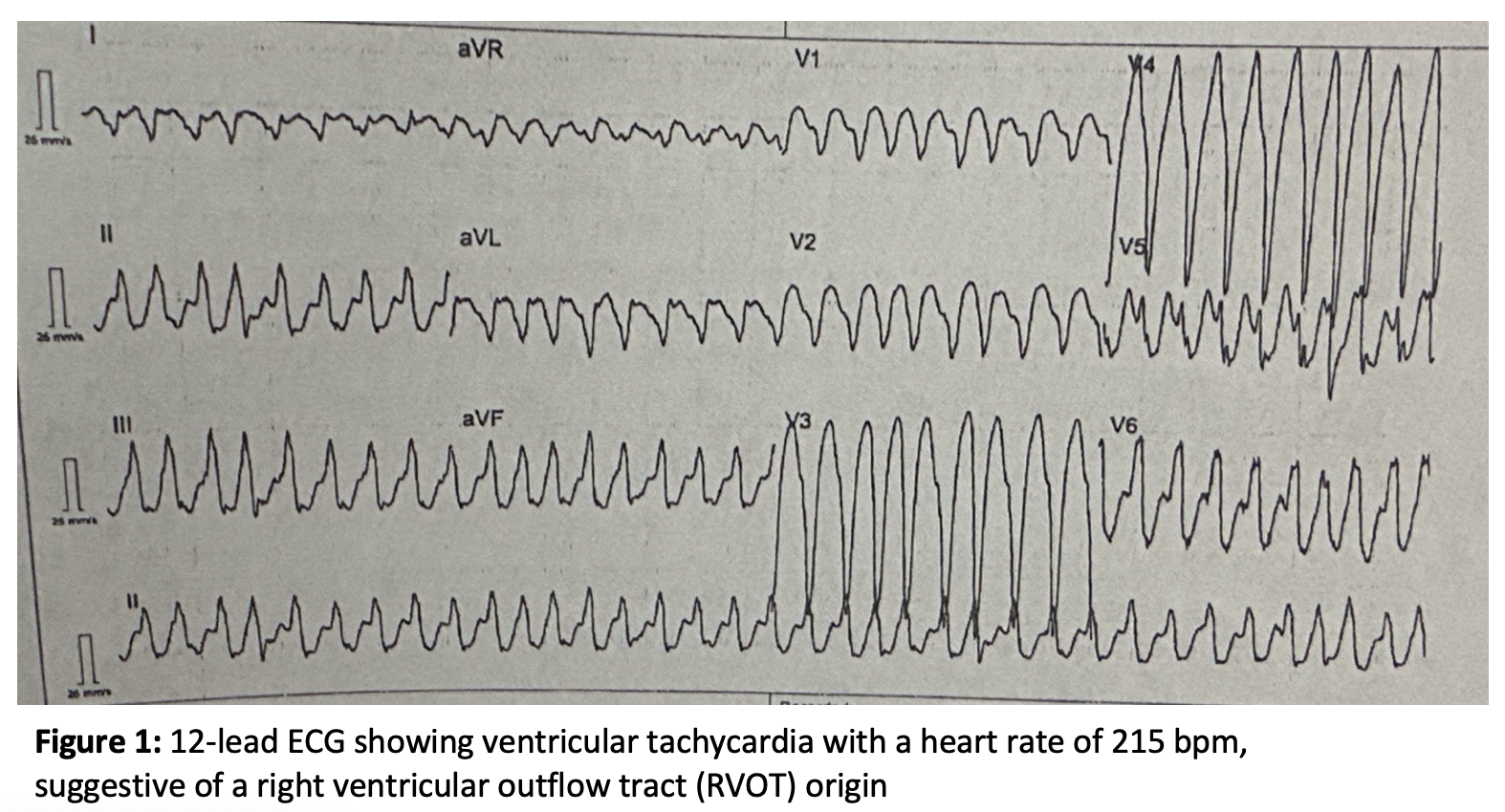
A 50-year-old caucasian patient was admitted with palpitations and angina. Initial Apple Watch ECG showed sustained VT (~215 bpm), later confirmed on 12-lead ECG (suggestive of RVOT origin), which was terminated by electrical cardioversion (Figure 1).
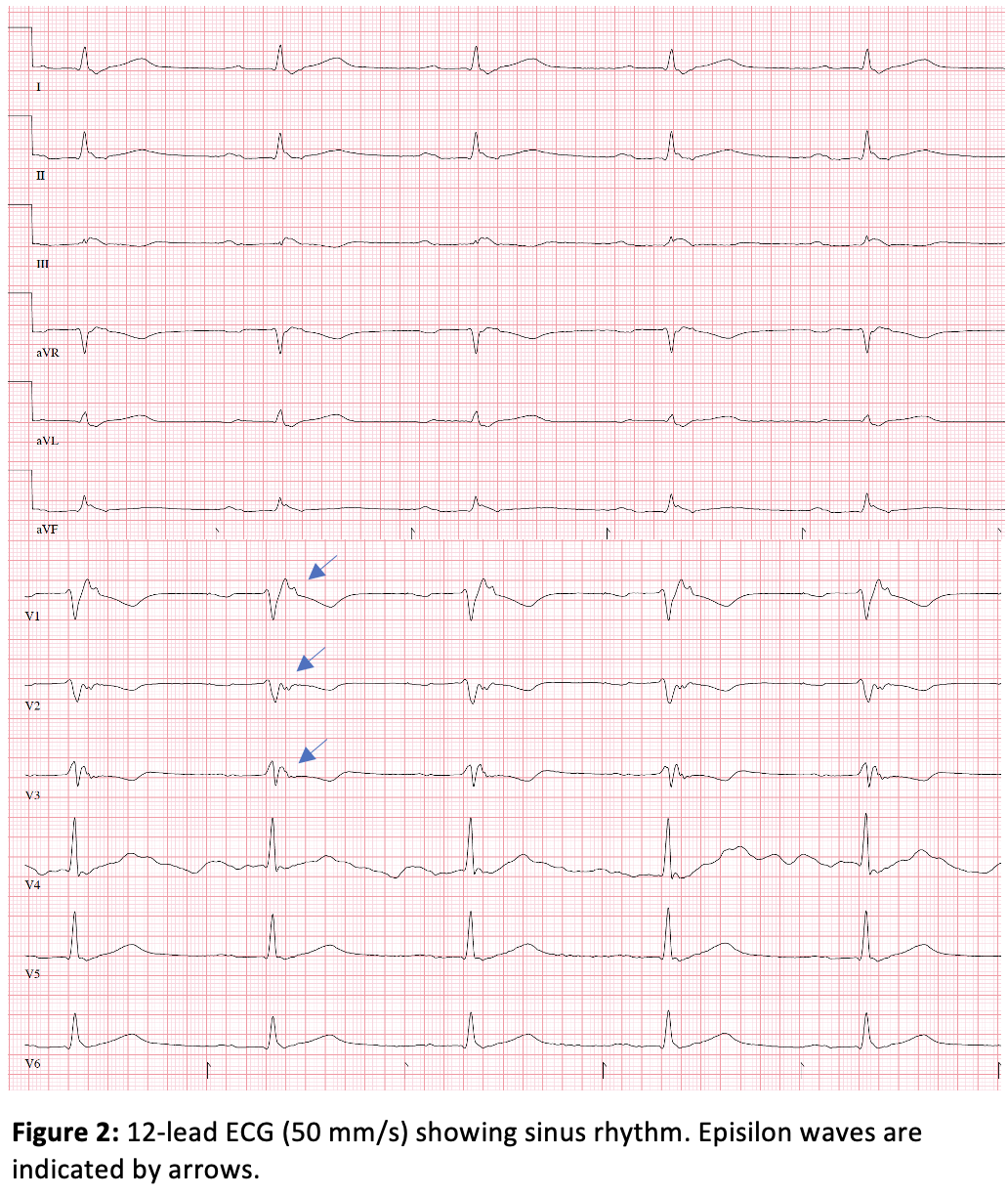
Baseline ECG in sinus rhythm showed first-degree AV block, right bundle branch block, T-wave inversion and prominent epsilon waves in V1-3 (Figure 2). Transthoracic echocardiography revealed preserved left ventricular function and moderately reduced right ventricular function with right ventricular dilatation, raising suspicion of ARVC.
Coronary artery disease was excluded by CT angiography. Cardiac MRI demonstrated biventricular late gadolinium enhancement (LG) with right ventricular dominance, however with atypical abnormalities not fully consistent with classical ARVC. An 18F-FDG PET/CT revealed cardiac sarcoidosis with atypical FDG uptake and active nodal and extranodal involvement, including the liver, spleen, bones, and lungs. In addition, the diagnosis of cardiac sarcoidosis was supported by histological findings from a lymph node biopsy.
For secondary prevention, a dual-chamber ICD was implanted and immunosuppressive therapy initiated. Two months later, the patient experienced recurrent VT and ventricular fibrillation with appropriate ICD therapies. Repeat PET-CT showed no active inflammation, suggesting a scar-related arrhythmogenic substrate. Despite amiodarone therapy, recurrent VT persisted, leading to referral for catheter ablation in our center.
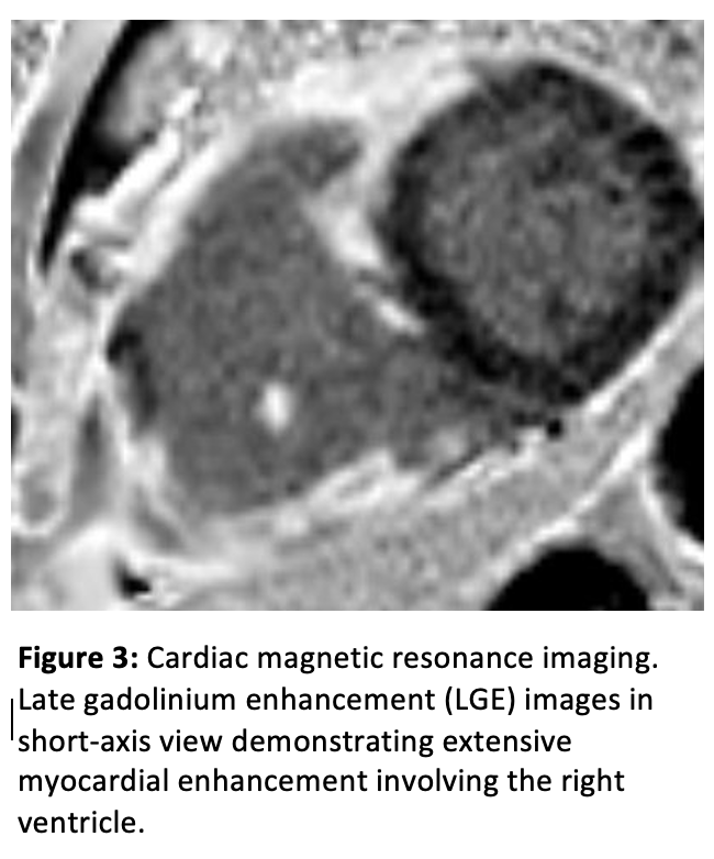
Repeat cardiac MRI showed extensive LGE involving the right ventricle (basal to apical), septum, and inferolateral left ventricle (Figure 3).
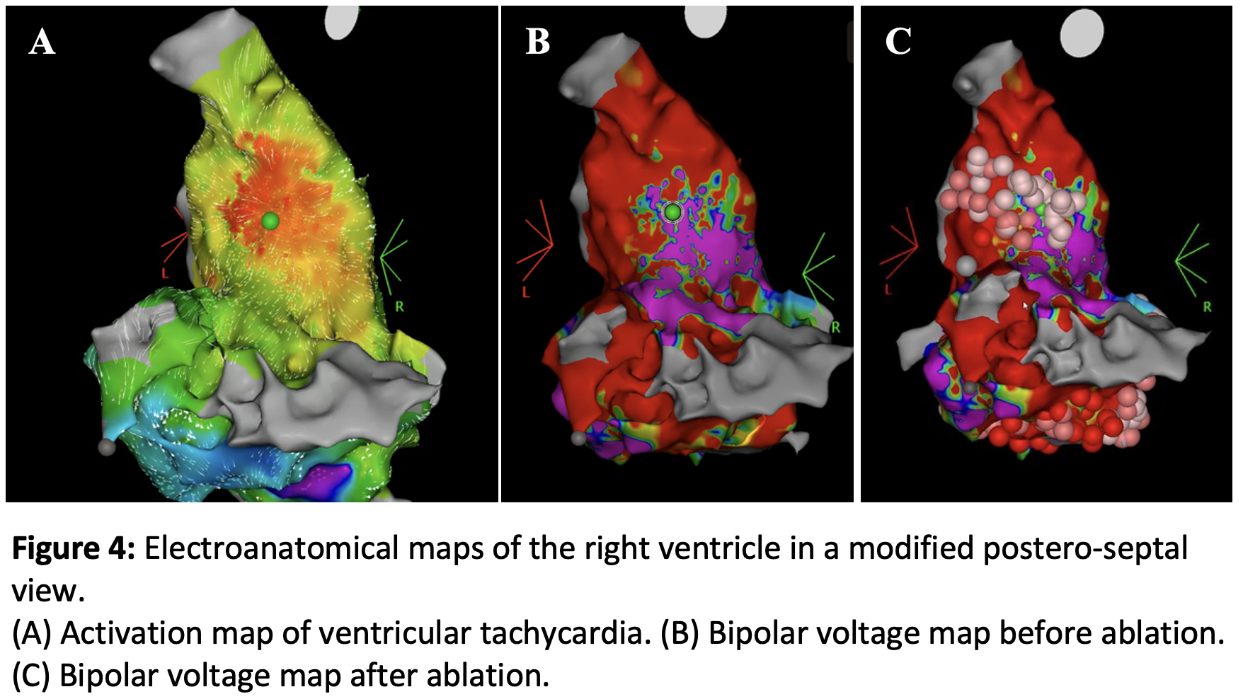
VT ablation was performed under Impella-supported hemodynamic assistance and deep sedation. High-density electroanatomical mapping revealed extensive low-voltage areas in the RVOT, septum, and inferior right ventricle with late potentials and fractionated electrograms. A spontaneous VT (TCL 350 ms) enabled activation mapping, consistent with an origin from the posterior RVOT (Figure 4 A, B). Substrate modification was performed (Figure 4C). A second VT (TCL 400 ms) was inducible, with activation mapping indicating an anteroseptal RVOT origin, and was terminated by ablation. Then, no VT was inducible. No complications occurred.
Conclusion
Epsilon waves are not specific for ARVC and may also occur in systemic inflammatory diseases with predominant right ventricular involvement, such as cardiac sarcoidosis. This case highlights the risk of misdiagnosis and the importance of multimodality imaging for identifying underlying systemic disease.
Management requires a combined approach including immunosuppressive therapy, antiarrhythmic drugs, device implantation, and catheter ablation in selected patients. In hemodynamically unstable VT, mechanical circulatory support may facilitate mapping and ablation. Despite advanced therapy, prognosis may remain limited, and follow-up is essential.