Background:
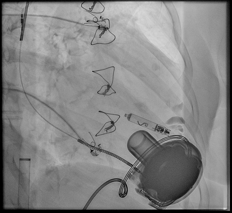
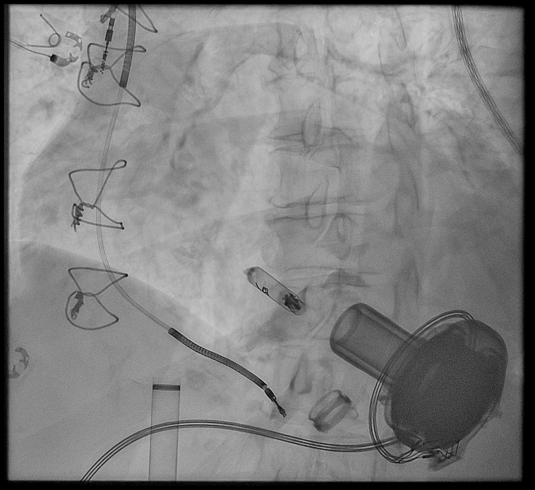
A 73-year-old woman with a left ventricular assist device (Medtronic HeartWare) as "destination therapy" for terminal, post-inflammatory dilated cardiomyopathy (post-Coxsackie myocarditis, 1986) and severely reduced left ventricular systolic function presented with recurrent slow ventricular tachycardias (slow VTs, 120–160/min). The patient had a history of recurrent infections of previous ICD systems, necessitating explantation. The option of a subcutaneous ICD (sICD) was considered; however, due to the predominance of slow VTs requiring antitachycardia pacing (ATP) and the absence of syncope – likely due to the support of the implanted LVAD – a approach using maximal antiarrhythmic therapy was maintained.
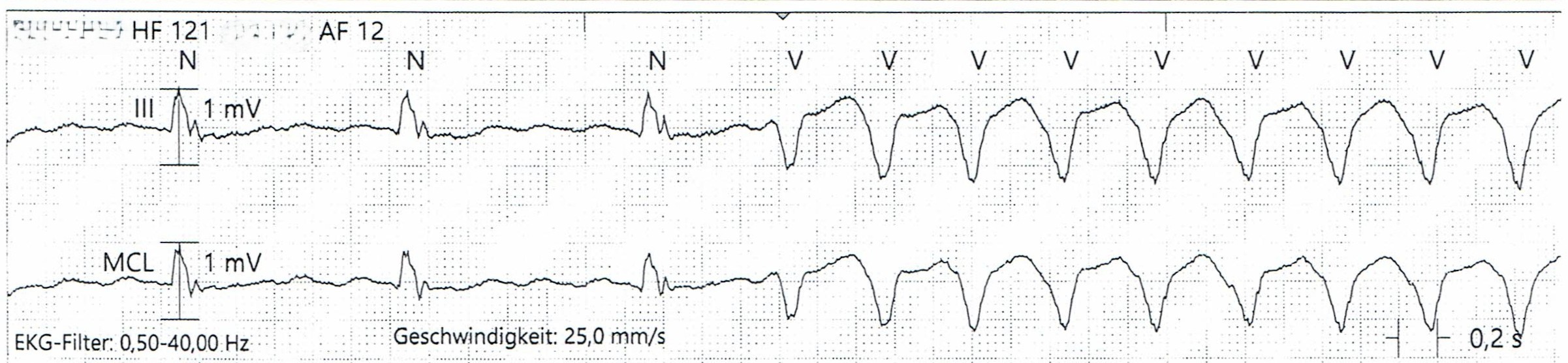
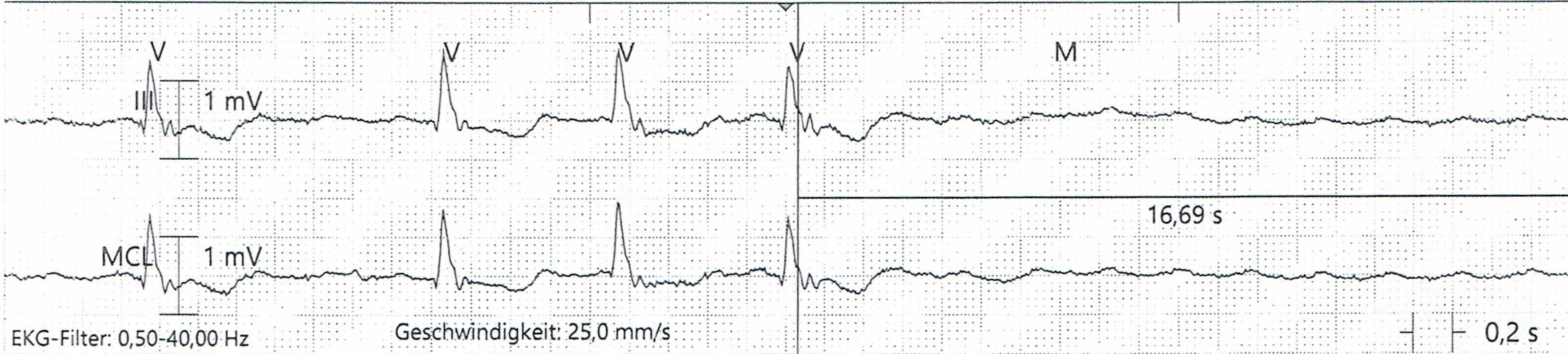
Under this treatment regimen, telemetry revealed no further ventricular arrhythmias but intermittent high-grade atrioventricular conduction disturbances with long pauses (up to 16 seconds). A ventricular stimulation was indicated to prevent symptomatic bradycardia, particularly given the lack of device-supported right ventricular function.
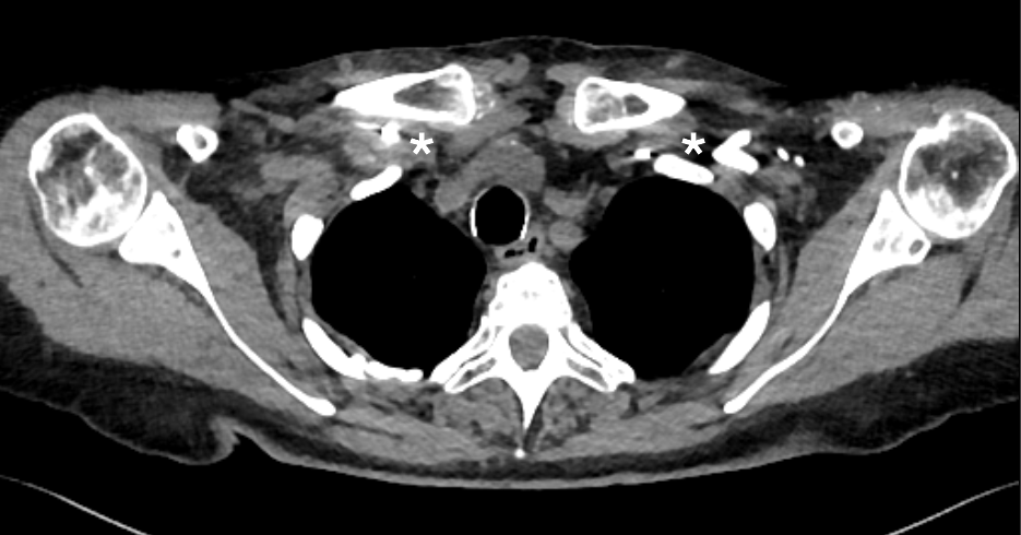
The situation was complicated by a thrombotic occlusions of the subclavian veins bilaterally and the superior vena cava, despite strict anticoagulation. Therefore the guideline directed indication for the implantation of a leadless pacemaker was established.
Methods:
Due to expected anatomical conditions, a system with active fixation was chosen (AVEIR™ VR). The interrogation of the AVEIR™ system is performed conductively via an electric field, which is detected by ECG electrodes. This battery saving communication is susceptible to external electrical signals (f.e. EP lab equipment, external defibrillator…) but necessary for the device implantation. The device can only be interrogated when it is in contact with blood. So the challenges of this procedure consisted of establishing the communication with the LCP as well as the anatomical conditions.
Results:
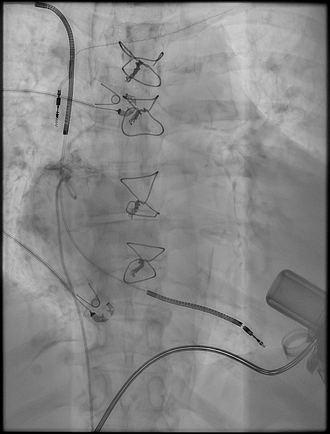
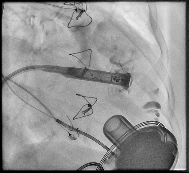
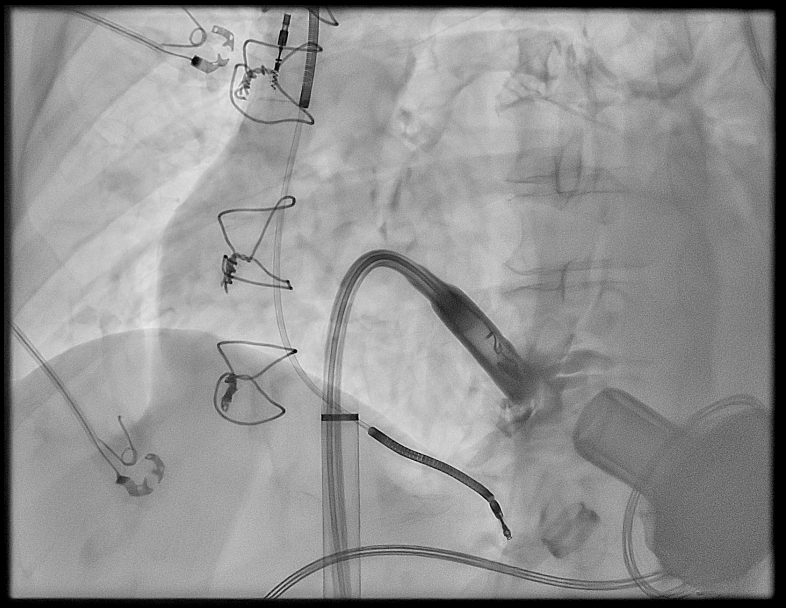
The leadless AVEIR™ VR pacemaker was successfully implanted using standard techniques under analgosedation. The right ventricular anatomy was visualized using a pigtail catheter. After successive predilation, a 25-French sheath was introduced, and the AVEIR™ VR LP was delivered to the anteroseptal region using the delivery catheter. The conductive communication was successfully established without any limitations. The device was repositioned twice due to altered anatomy following multiple prior cardiac surgeries (specifically LVAD implantation) with subsequent mapping. Final placement yielded good and stable impedance and stimulation threshold values in the anteroseptal position.
Discussion/ Conclusion:
Ventricular arrhythmias and high-grade AV-block compromises right ventricular function in patients with left ventricular assist device. This case describes the implantation of a leadless AVEIR™ VR LP pacemaker in an adult LVAD patient for terminal post-inflammatory dilated cardiomyopathy, permanent atrial fibrillation and third-degree AV block, aimed at ensuring maximal antiarrhythmic therapy for ventricular arrhythmias in the setting of limited thoracic access due to venous thrombotic occlusion. The technical prerequisites, feasibility, and safety were demonstrated despite anatomical and telemetric challenges.