Introduction:
Arrhythmogenic Right Ventricular Cardiomyopathy (ARVC) is a rare inherited cardiomyopathy associated with ventricular arrhythmias and an increased risk of sudden cardiac death, particularly in young individuals. Implantable cardioverter- defibrillator (ICD) therapy is central for the management of ARVC. However, device selection in pediatric patients remains challenging due to their small size and long-term complications of transvenous systems.
Case summary:
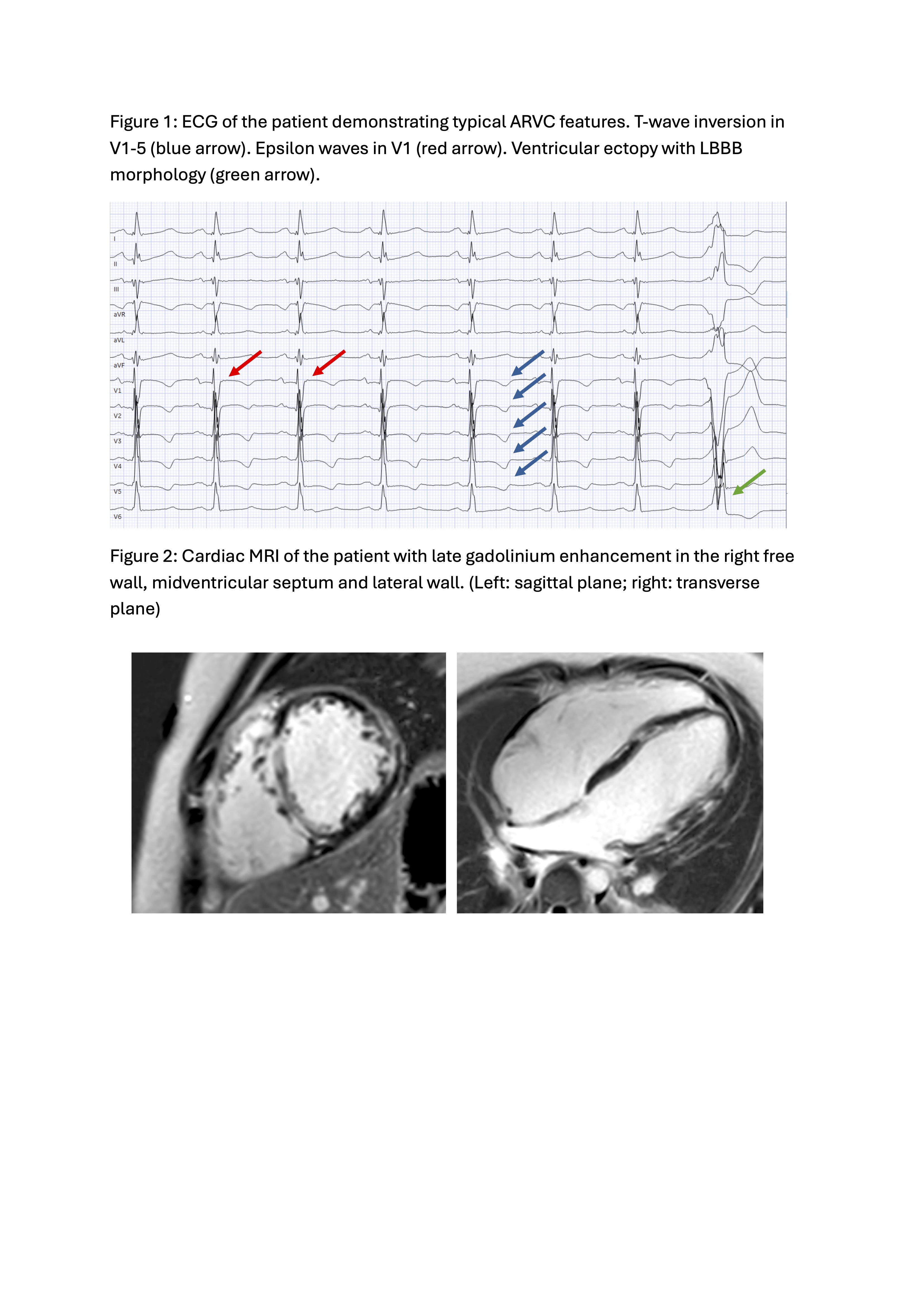
We report the history of an 8-year-old child who suPered from out-of-hospital cardiac arrest with ventricular fibrillation and was successfully resuscitated. During hospitalization, recurrent ventricular tachycardia occurred, degenerating into ventricular fibrillation, requiring repeated resuscitation. ARVC was diagnosed based on fulfillment of 3 major criteria of the ARVC Task Force criteria. The 12 lead ECG demonstrated epsilon waves in V1 (Figure 1) and T-wave inversion in V1-5 and genetic testing revealed a pathogenic mutation in the PKP2 gene. Magnetic resonance imaging revealed impaired biventricular systolic function and pronounced late gadolinium enhancement in both ventricles (Figure 2).
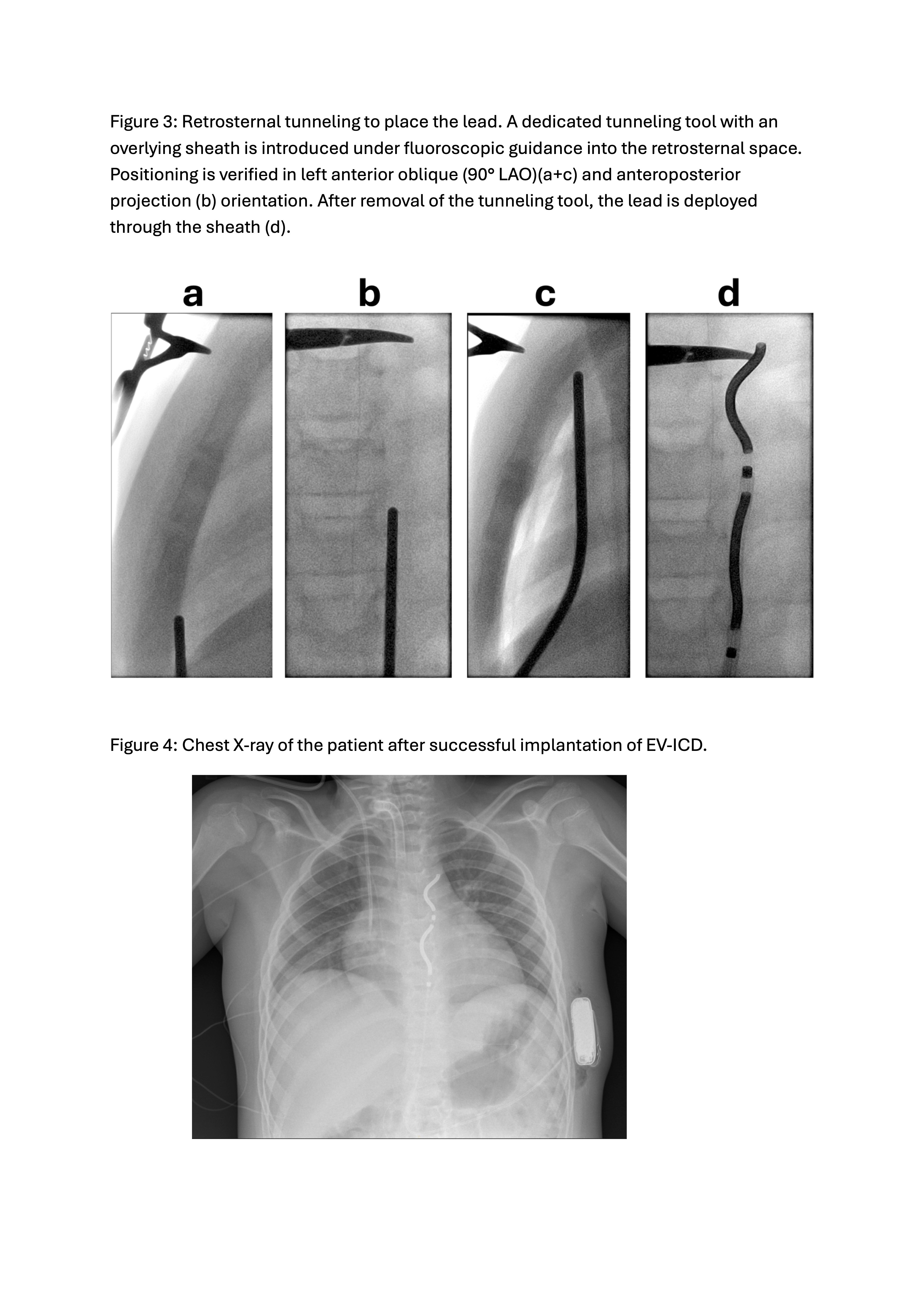
Indication for secondary preventive ICD implantation was established. Considering the patient’s young age, small body size (42kg and 140cm) and the need to avoid long-term intravascular lead complications, the decision for implanting an extravascular ICD (EV- ICD) was made. Preoperative thoracic computed tomography excluded thoracic deformities and revealed a sternal length of 14cm. EV-ICD was implanted under general anesthesia with fluoroscopic guidance. Prior to the procedure, anatomical landmarks including the xiphoid process, left parasternal border, and the planned generator position along the left midaxillary line were marked. A subxiphoid incision was made, followed by blunt dissection and retrosternal tunneling along the left parasternal border to position the lead (Figure 3). After confirming adequate sensing without P-wave oversensing, the lead was secured to the rectus fascia, tunneled and connected to the generator in the subcutaneous generator pocket. Intraoperative defibrillation testing demonstrated reliable detection and ePective defibrillation with a single 30 J shock. Postoperative chest x-ray revealed adequate device position (Figure 4) and device interrogation showed stable sensing across multiple vectors and body positions. During short term follow-up the patient presented with stable condition. Neurological status showed improvement compared to the immediate post-resuscitation period.
Discussion:
This case highlights an aggressive arrhythmic phenotype of ARVC in a pediatric patient and reports successful EV-ICD implantation as a promising non-transvenous option for young patients needing an ICD.