Introduction
Reliable atrial sensing and capture are essential for dual-chamber pacing. Exit block is typically attributed to technical issues such as lead dislodgement or dysfunction, and evaluation focuses on lead integrity and device parameters. However, many patients requiring atrial pacing have a history of atrial arrhythmias and prior ablation, associated with progressive atrial remodeling. Fibrosis and conduction abnormalities can alter atrial electrophysiology and, in advanced stages, lead to electrical compartmentalization.
Case description
A 51-year-old woman was referred for ablation of atrial tachycardia (AT). She had previously undergone pulmonary vein isolation and cavotricuspid isthmus ablation at another institution and had received a dual-chamber pacemaker for sinus arrest and complete atrioventricular block. Structural heart disease was not present.
Preprocedural device interrogation (in sinus arrest) revealed exit block of the right atrial (RA) lead, despite stable lead impedance. Chest X-ray excluded relevant lead dislodgement.
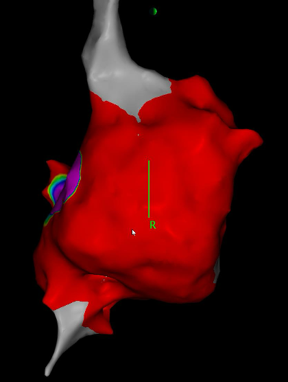
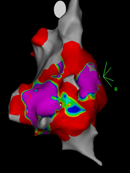
Given the absence of lead dislodgement, we proceeded as initially planned with left atrial AT ablation including anterior and roof lines. To assess the RA substrate prior to considering lead revision, a high-density voltage map of the RA was acquired during the procedure. Owing to permanent sinus arrest, mapping was performed under coronary sinus pacing using a multipolar mapping catheter. The voltage map revealed extensive low-voltage areas (<0.5 mV), consistent with advanced atrial scarring (Fig. 1). The scarring resulted in electrical dissociation of the lateral RA (Fig. 2), thereby providing a mechanistic explanation for the observed RA lead exit block.
A small septal region located posterosuperior to the coronary sinus (CS) ostium demonstrated preserved conduction to the left atrium. Based on these findings, RA lead revision was planned, targeting lead placement in this area.
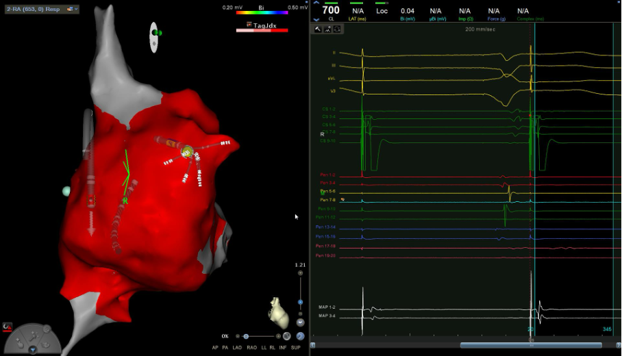
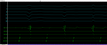
The procedure was initiated with placement of a decapolar catheter in the CS via femoral access to monitor atrial activation. Subsequently, the axillary vein was accessed using an ultrasound-guided approach. Initial attempts to position the RA lead using a His catheter were unsuccessful. Therefore, a steerable sheath was employed, allowing stable lead deployment in the posterosuperior septal region adjacent to the CS ostium. A lumenless lead was used. Surface ECG did not demonstrate discernible P waves. However, effective atrial capture was confirmed via CS recordings (Fig. 3), indicating restoration of functional atrial activation and a successful procedural outcome.
Discussion
RA lead exit block is usually attributed to technical causes. This case illustrates advanced atrial scarring as an alternative mechanism. Atrial remodelling and previous ablation may electrically isolate atrial regions such that local capture does not result in effective atrial activation. Consequently, exit block can occur despite intact lead function. In this case, voltage mapping identified lateral RA dissociation, whereas a septal region retained conduction, enabling successful lead implantation. However, the absence of discernible P waves raises uncertainty as to whether this translated into meaningful clinical benefit. Overall, this case highlights that exit block should not be equated with lead failure alone and that electroanatomical mapping may guide lead revision in patients with extensive atrial scarring.