Background
Calcified coronary lesions are increasingly encountered during percutaneous coronary intervention (PCI) and remain among the most challenging lesion subsets because of their morphological complexity. At the same time, there is a growing focus on restoring physiological vasomotion and vascular remodeling in the context of PCI.
Case Summary
We present a case series of three patients with severely calcified coronary lesions undergoing PCI. Lesion preparation was performed using contemporary techniques, including rotational atherectomy (RA) in two of three cases, followed by implantation of a bioadaptor – a sirolimus-eluting cobalt-chromium drug-eluting coronary hybrid stent system (Elixir Medical, Milpitas, CA, USA). In one case, a super high-pressure OPN balloon was used for post-dilatation. Six-month follow-up angiography demonstrated favourable outcomes with sustained vessel patency in all cases.
Case 1: A 48-year-old male presented with chest pain. Angiography showed severe distal left circumflex artery (LCX) calcification. Optical coherence tomography (OCT)-guided PCI showed a 190° calcium arc (length: 21 mm, minimal lumen area (MLA): 2.0 mm²). Following non-compliant (NC) balloon preparation with good balloon expansion and a favourable angiographic result, a 3.0 × 23 mm bioadaptor was deployed. Underexpansion was corrected using a super high-pressure OPN balloon at 30 atm (good angiographic result, >80% expansion, minimal stent area (MSA): 5.94 mm²). At 6-month angiographic follow-up, results were favourable and the patient was asymptomatic.
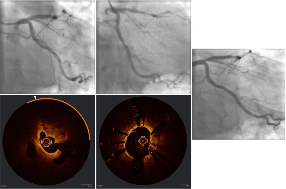 Case 2:
 A 72-year-old male presented with reduced exercise tolerance. Angiography showed severe proximal left anterior descending artery (LAD) stenosis. OCT revealed a 180° calcium arc (length: 19 mm, MLA: 1.19 mm²). Lesion preparation required RA and NC balloon predilation. A 3.5 × 23 mm bioadaptor was deployed and post-dilated (good angiographic result, proximal in stent: >80% expansion, MSA: 10.44 mm², distal in the stent: >80% expansion, MSA 7.81 mm²). At 6-month angiographic follow-up, results remained favourable and the patient was symptom-free.
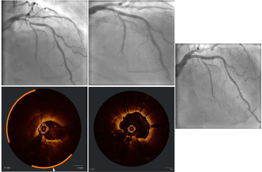 Case 3:
 A 71-year-old female presented with progressive dyspnea. Angiography confirmed severe proximal LAD stenosis. OCT showed a 190° calcium arc (length: 22 mm, MLA: 2.77 mm²). RA followed by cutting balloon and NC balloon preparation was performed. A 3.5 × 23 mm bioadaptor was deployed and post-dilated (good angiographic result, proximal in stent: >80% expansion, MSA: 10.56 mm², distal in the stent: >80% expansion, MSA 7.85 mm²). At 6-month angiographic follow-up, the patient was asymptomatic with a favourable result.
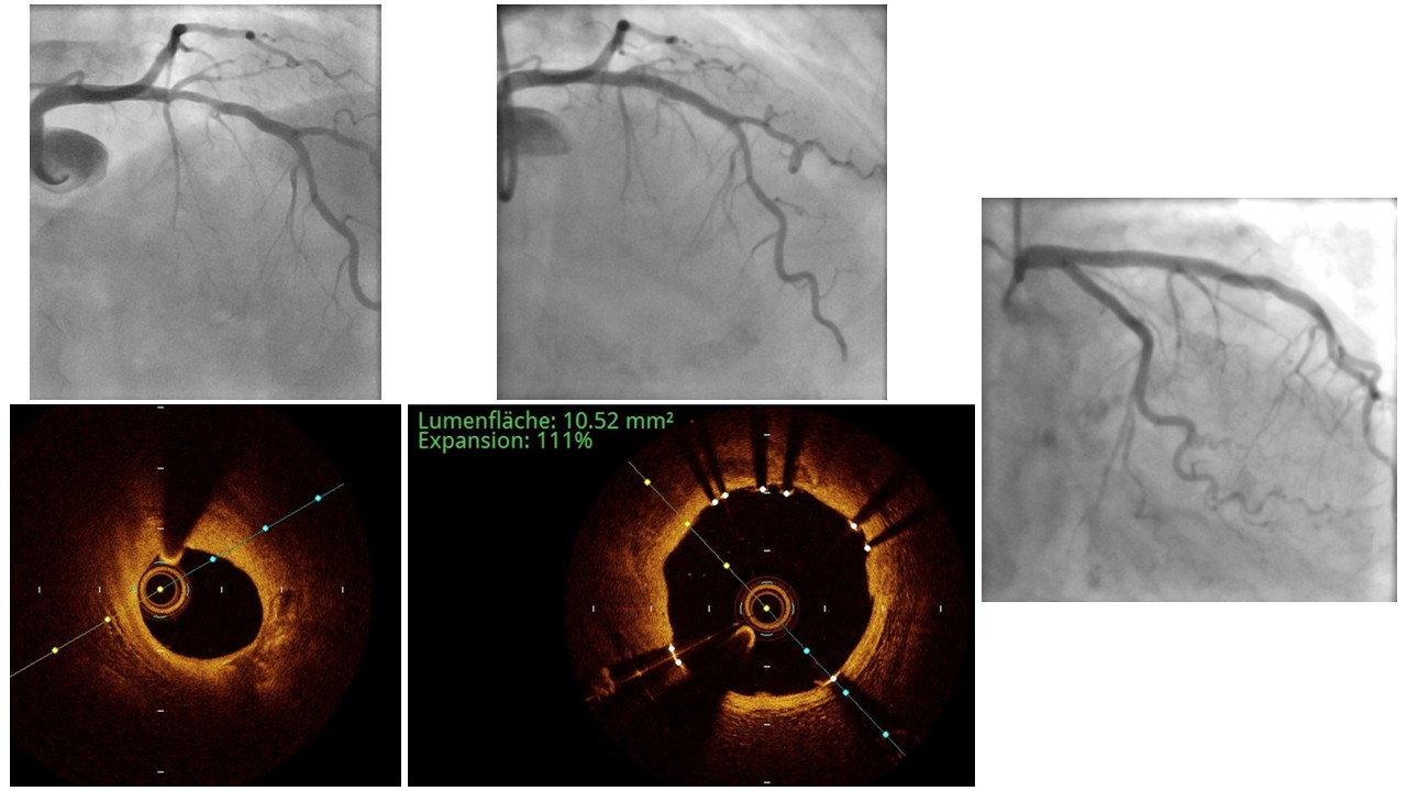
Conclusion
To the best of our knowledge, this is the first case series reporting the use of a bioadaptor in heavily calcified coronary lesions, with a super high-pressure OPN balloon used in one case and RA performed in two of three cases. Adequate lesion preparation, especially by means of RA, along with intravascular imaging guidance, is essential in the treatment of calcified coronary stenoses, given its impact on procedural success and clinical outcomes.
Key learning points:
- The use of a bioadaptor in calcified coronary lesions is feasible and safe.
- Optimal lesion preparation in calcified coronary lesions, especially using RA, and the use of intravascular imaging to guide the procedure are essential for favourable treatment outcomes.



