Background: Coronary obstruction is a rare but potentially fatal complication of transcatheter aortic valve implantation (TAVI). The risk is markedly increased in patients with adverse aortic root anatomy and congenital coronary anomalies. We report a case of successful coronary protection during TAVI in a patient with a single coronary artery and multiple predictors of coronary obstruction.
Case Presentation: An 86-year-old woman with symptomatic severe aortic stenosis (NYHA III) was referred for transfemoral TAVI. Her medical history included heart failure with reduced ejection fraction secondary to valvular cardiomyopathy, arterial hypertension, dyslipidemia, peripheral arterial disease, and recent PCI of the LAD with implantation of three drug-eluting stents.
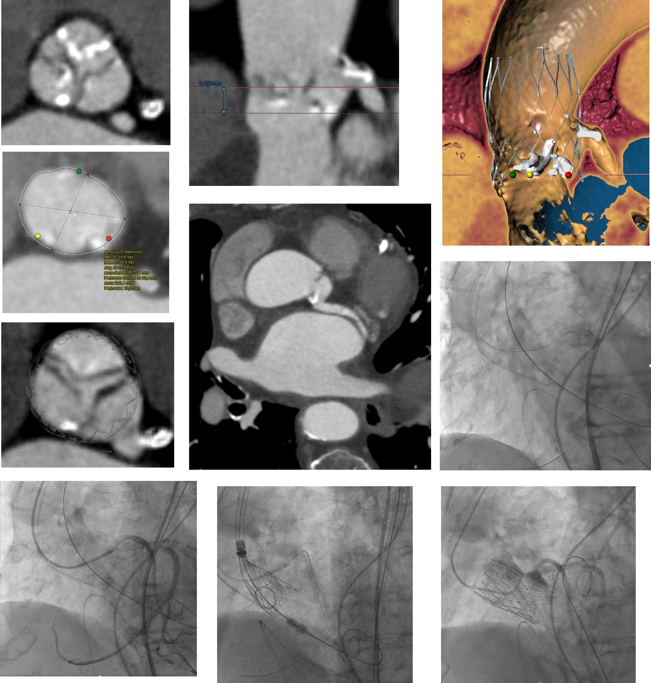
Echocardiography demonstrated severe high-gradient aortic stenosis (Vmax 4.92 m/s, mean gradient 58.9 mmHg, AVA 0.42 cm², stroke volume index 33.9 ml/m²). Preprocedural CT revealed several established risk factors for coronary obstruction, including a left main coronary height of 7 mm, sinus of Valsalva diameter of 25.9 mm, and a virtual valve-to-coronary distance (VTC) of 2.5 mm. Annular measurements showed an area of 383 mm² and a perimeter of 70.5 mm.
Coronary angiography demonstrated a congenital single coronary artery arising from the left coronary sinus, with the right coronary artery originating from the distal circumflex artery, rendering the entire myocardium dependent on the left coronary system.
Given the small annular dimensions, a self-expanding supra-annular system was selected. In addition, in view of the prohibitive risk of coronary obstruction, this platform was preferred to allow controlled deployment in a complex coronary anatomy. Ultimately, a 27-mm self-expanding supra-annular Allegra valve was implanted.
Following transfemoral access and temporary pacing lead placement, balloon aortic valvuloplasty with a 20-mm balloon was performed under rapid pacing. During inflation, transient left main compromise resulted in ventricular fibrillation and hemodynamic collapse. Immediate defibrillation restored stable circulation.
The left main coronary artery was subsequently engaged with an EBU 3.75 guiding catheter. Protective guidewires were advanced into the LAD and LCX, and a balloon was positioned in the LCX for potential bailout intervention. Thereafter, the 27-mm Allegra valve was successfully implanted. Final angiography confirmed preserved coronary perfusion, optimal valve position, and absence of significant paravalvular regurgitation.
Discussion: This case illustrates the cumulative impact of multiple anatomical risk factors for coronary obstruction during TAVI. The combination of extremely low coronary height, narrow sinus of Valsalva, critical VTC and a single coronary artery created an exceptional high-risk setting. Ventricular fibrillation during balloon valvuloplasty represented an early manifestation of transient left main obstruction and prompted immediate coronary protection.
Conclusion: Patients with a single coronary artery and adverse aortic root anatomy represent an exceptionally high-risk subgroup for coronary obstruction during TAVI. Careful CT-based planning, appropriate valve selection, and rapid implementation of coronary protection strategies are essential to ensure procedural success.
Keywords: TAVI, single coronary artery, coronary obstruction, coronary protection, ventricular fibrillation, Allegra valve.