Patient PresentationAn 82-year-old man presented to our clinic with severe dyspnea (NYHA IV) and hypoxemia three months after heterotopic bicaval valve implantation for recurrent massive tricuspid regurgitation. Oxygen saturation was 68% on room air and 75% despite 8 L/min supplemental oxygen.
His history included multiple structural interventions: two prior tricuspid transcatheter edge-to-edge repairs (T-TEER) for recurrent severe tricuspid regurgitation and one mitral TEER (M-TEER) for severe mitral regurgitation. Despite these therapies, he developed recurrent right-sided heart failure with peripheral edema. He also developed recurrent large pericardial effusions requiring repeated drainage. Cytological analysis showed a protein rich and hemorrhagic pericardial effusion without evidence of malignancy or active inflammatory disease. Comorbidities included persistent atrial fibrillation, hypertension, chronic kidney disease, and non-obstructive coronary atherosclerosis. After bicaval valve implantation, he initially improved and was discharged on guideline-directed medical therapy.
Initial Work-UpECG showed atrial fibrillation (59 bpm) with right bundle branch block. Laboratory tests revealed elevated troponin T (45.7 ng/L) and NT-proBNP (177 pmol/L). Chest X-ray presented no pulmonary infiltrates.
Transthoracic echocardiography showed a large circumferential pericardial effusion (up to 3.4 cm), mildly reduced LV function (EF 50%), and mildly impaired RV function with reduced free-wall strain. Residual moderate mitral regurgitation and persistent massive tricuspid regurgitation were present. Bicaval valve prostheses were correctly positioned and functional. Color Doppler revealed a persistent iatrogenic atrial septal defect (iASD) related to prior M-TEER.
Diagnosis and ManagementPericardial drainage was performed, draining 2000 mL of serous fluid, but hypoxemia persisted with SpO₂ ~70%, indicating an alternative explanation. Further evaluation with transesophageal echocardiography revealed a 12×6 mm iASD with bidirectional flow. Right heart catheterization showed pulmonary artery pressure of 46/7 (23) mmHg and prominent biatrial v-waves, with higher right atrial pressures. Near equalization of atrial pressures across the ASD suggested a significant interatrial communication.
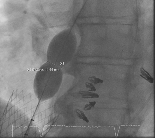
Given severe hypoxemia and suspected right-to-left shunting, balloon test occlusion of the defect was performed. Oxygenation improved without significant rise in LVEDP (10–12 mmHg), indicating good tolerance. Balloon sizing measured an 11 mm defect. Hemodynamic assessment confirmed a bidirectional shunt with predominant right-to-left flow.
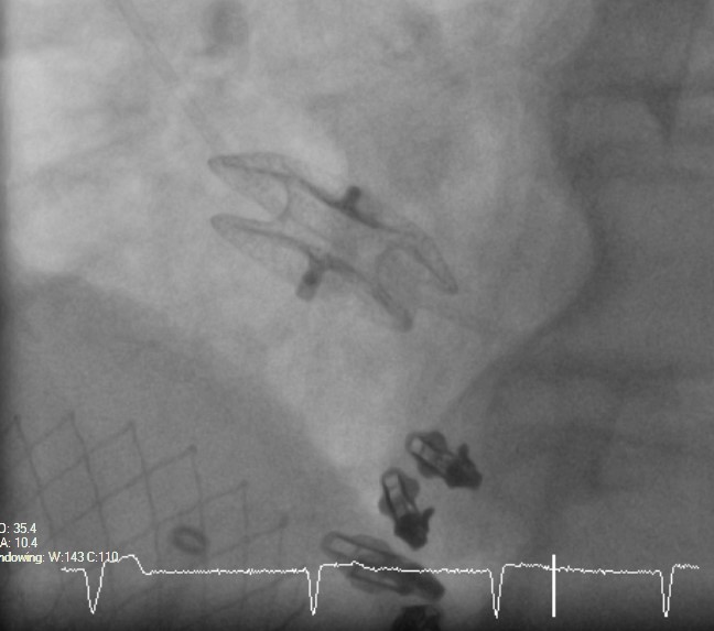
The defect was closed with a 14-mm occluder device. Oxygen saturation improved from 89% on high-flow oxygen to 98% during the procedure.
Follow-UpThe patient remained stable without supplemental oxygen. Echocardiography showed correct device position, minimal residual shunt, preserved LV function, and stable mildly reduced RV function with reduction of pericardial effusion.
At 1- and 3-month follow-up, he was asymptomatic with oxygen saturation 97–98% on room air. Repeat TEE confirmed stable occluder position with only minimal residual contrast passage.
ConclusionPersistent previously silent iASD can become clinically significant after bicaval valve implantation due to increase in right atrial pressure.Therefore, percutaneous closure of the defect should be considered before heterotrophic bicaval valve implantation.