Background: A pancreatopericardial fistula is an exceptionally rare, life-threatening complication of chronic pancreatitis resulting from pancreatic duct disruption. Due to non-specific symptoms and standard imaging limitations, establishing an accurate diagnosis remains highly challenging.
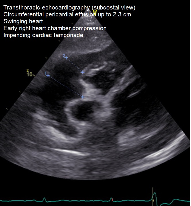
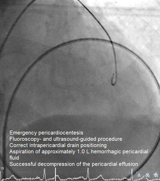
Case Presentation: A 60-year-old male with known chronic pancreatitis presented with progressive dyspnea. Transthoracic echocardiography (TTE) demonstrated a preserved LVEF without regional wall motion abnormalities, but revealed a massive 2.3 cm circumferential pericardial effusion with a swinging-heart phenomenon and early right-sided chamber compression, indicating impending cardiac tamponade. Urgent pericardiocentesis was performed successfully. Unexpectedly, fluid analysis revealed a profound exsufflation of pancreatic enzymes with a markedly elevated lipase level (>5,000 U/L), suggesting an extra-cardiac origin.
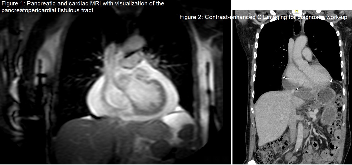
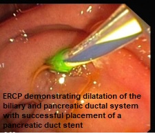
To rule out primary cardiovascular etiologies, an extensive cardiac workup was initiated. Technetium-99m bone scintigraphy demonstrated no pathological myocardial tracer uptake, effectively excluding cardiac amyloidosis and other systemic storage diseases. Subsequent diagnostic workup via ultrasound, contrast-enhanced CT, thoracic imaging, and ERCP indicated pancreatic duct pathology but failed to visualize the definitive fistulous communication. The anatomical tract was ultimately delineated using dedicated MRI and MRCP, confirming a complex pancreatopericardial fistula arising from the pancreatic bed, coursing through a short transhepatic segment, and extending via the caval foramen directly into the pericardium.
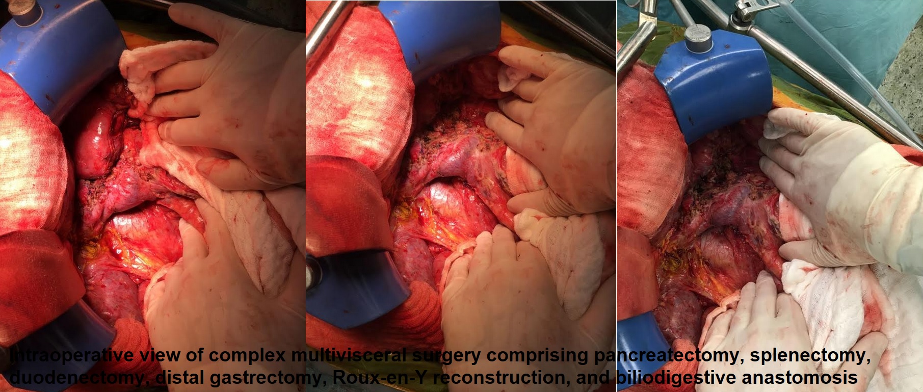
Advanced morphological evaluation revealed extensive parenchymal changes, including an inflammatory pseudotumor measuring >3 cm in the pancreatic head and a 4 cm mass in the pancreatic tail (cauda). Fine-needle aspiration (FNA) of these lesions confirmed high local concentrations of pancreatic enzymes. Histopathological and cytological evaluation yielded morphological features diagnostic of chronic pancreatitis, with no evidence of IgG4-associated disease. Given the recurrence of symptomatic effusions refractory to conservative regimens, combined with the extensive bilateral masses, a clear indication for surgical intervention via a pylorus-preserving pancreaticoduodenectomy (PPPD) was established. The patient underwent a complex, 9-hour procedure comprising extensive adhesiolysis, PPPD, splenectomy, duodenectomy, distal gastric resection, Roux-en-Y reconstruction, and bilioenteric anastomosis.
Conclusion: Markedly elevated pancreatic enzymes within pericardial fluid must prompt targeted gastrointestinal evaluation. In occult fistulae, MRI/MRCP offers superior diagnostic sensitivity compared to CT/ERCP. Refractory cases associated with advanced parenchymal disease and large inflammatory pseudotumors (>3 cm in the head, 4 cm in the tail) without IgG4 association necessitate timely multidisciplinary management and complex surgery, such as a PPPD, as a definitive curative approach.