Background
Cardiac involvement in Burkitt lymphoma is exceedingly rare and usually reflects advanced systemic disease with secondary myocardial infiltration rather than a primary cardiac malignancy. Due to often nonspecific clinical presentation, diagnosis remains challenging. In this setting, cardiac magnetic resonance imaging (CMR) plays a pivotal role by enabling comprehensive tissue characterization and providing a form of “in vivo histology” through advanced contrast-enhanced imaging techniques.
Case Report
A 61-year-old man was admitted to the hematology-oncology intensive care unit with an unclear abdominal mass presenting with subileus symptoms and newly developed acute heart failure. He had a known HIV infection since 09/2018 (CDC stage C3; viral load 3 × 10⁵ copies/mL) and a history of Burkitt lymphoma diagnosed in 10/2018.
Biopsy of the abdominal mass showed no definite evidence of malignancy; however, extensive necrosis with residual CD20 positivity raised suspicion for recurrent lymphoma. Transthoracic echocardiography revealed a suspicious right ventricular mass associated with septal akinesia, suggestive of myocardial infiltration.
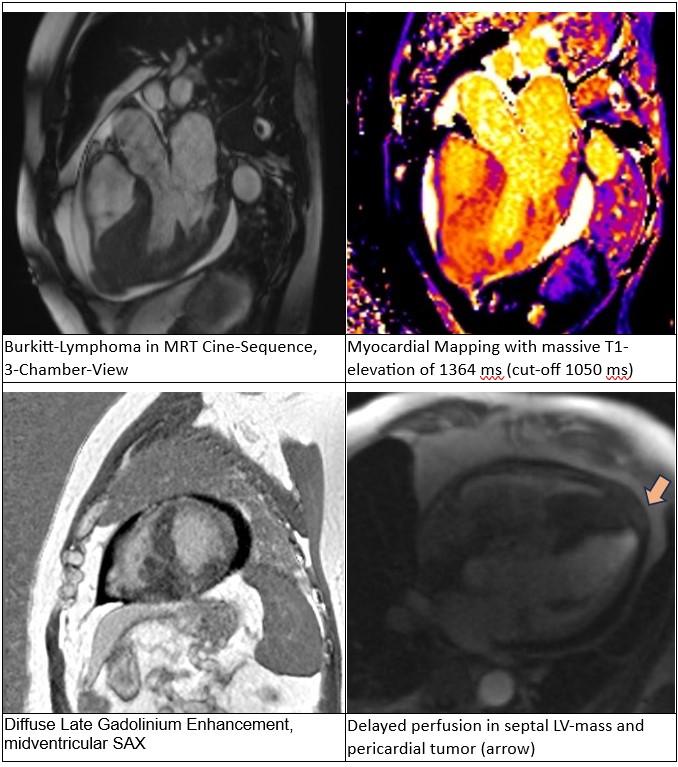
Cardiac MRI was performed for further tissue characterization and demonstrated a normal-sized left ventricle with moderately reduced systolic function, while the right ventricle was normal in size with preserved systolic function. A large infiltrative mass caused extensive myocardial thickening of the anteroseptal, inferoseptal and inferior left ventricular walls, extending into the right ventricular myocardium and papillary muscles. An additional pericardial lesion was identified at the left ventricular apex. The mass showed heterogeneous signal characteristics with diffuse late gadolinium enhancement, elevated native T1/T2 values, increased extracellular volume and delayed perfusion, consistent with a highly vascularized infiltrative tumor. Overall, findings were highly suggestive of recurrent Burkitt lymphoma with combined intramyocardial and epicardial involvement.
Twelve endomyocardial biopsies obtained from the right ventricular septum confirmed the diagnosis of Burkitt lymphoma. Histopathology demonstrated features of a high-grade B-cell lymphoma, while molecular genetic analyses indicated a de novo disease rather than relapse of the previously treated lymphoma. In accordance with German Multicenter Study Group for Adult Acute Lymphoblastic Leukemia recommendations, treatment with prednisolone and cyclophosphamide was initiated.
Fivedays later, the patient developed cardiogenic shock with pulmonary edema. Transthoracic echocardiography revealed a newly developed apical ventricular septal defect within the area of prior myocardial lymphoma infiltration, most likely therapy-associated. As neither surgical nor interventional repair was considered feasible, a palliative treatment strategy was pursued. The patient died two days later, four weeks after the onset of initial symptoms.
Conclusion
This case highlights the aggressive and fulminant nature of cardiac involvement in HIV-associated Burkitt lymphoma. CMR enabled comprehensive characterization of the infiltrative myocardial mass and played a crucial role in guiding the diagnostic pathway by providing noninvasive “in vivo histology.” Despite rapid interdisciplinary management, extensive myocardial infiltration was associated with a catastrophic clinical course and fatal outcome.