Background:
Adult patients with repaired Tetralogy of Fallot (ToF) are at increased risk of sustained ventricular tachycardia (VT). Catheter ablation can be challenging due to complex post-surgical substrates, altered cardiac anatomy, and atypical arrhythmogenic circuits. We report a case of recurrent VT in a patient with repaired ToF undergoing redo catheter ablation after failure of previous ablation procedures.
Case Presentation:
We report a 45-year-old woman with ToF, who underwent initial surgical repair in 1983 with subsequent surgical pulmonary valve replacement in 2013, followed by transcatheter pulmonary valve implantation for homograft degeneration in 2024. She presented with very frequent, highly symptomatic episodes of poorly tolerated monomorphic VT requiring ICD therapies with antitachycardia pacing.
The recurrent VT persisted despite antiarrhythmic therapy with sotalol and two previous VT ablation procedures with substrate modification of the anterolateral conduit and adjacent right ventricular outflow tract (RVOT) VT substrate.
The patient was referred for repeat electrophysiological study and ablation. Three-dimensional electroanatomical mapping of the right ventricle demonstrated a well-defined substrate region with numerous local abnormal ventricular activities (LAVAs) extending from the septal to the anterolateral RVOT in the setting of markedly distorted post-surgical anatomy and a severely rotated cardiac axis. Programmed ventricular stimulation reproducibly induced the clinical sustained monomorphic VT (TCL 320 ms).
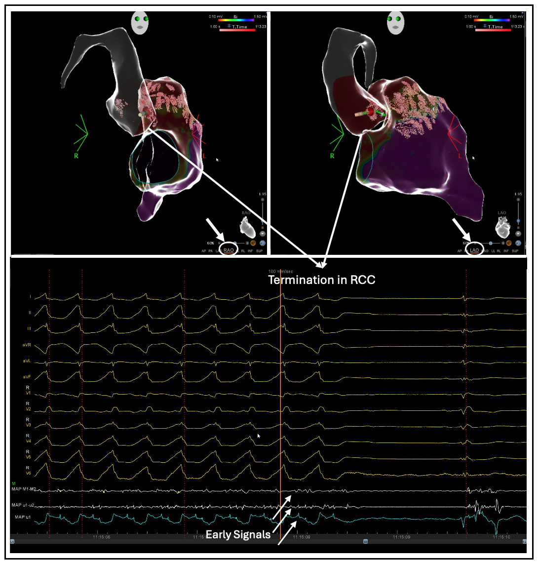
Activation mapping localized the earliest activation to the posteroseptal RVOT. Extensive substrate modification was initially performed, eliminating all identifiable LAVAs and fractionated electrograms within the RVOT. Following re-induction of VT, activation mapping raised suspicion of an epicardial component of the VT circuit, while pace mapping within the RVOT was non-diagnostic. Therefore, a retrograde transaortic approach was undertaken. Mapping of the aortic root identified the earliest activation within the right coronary cusp (RCC). Radiofrequency application at this site resulted in immediate and specific termination of the VT (Figure 1). VT was no longer inducible with programmed stimulation up to 330S4. Follow-up of more than four months demonstrated no VT recurrence.
Conclusion:
The persistence of inducible VT despite extensive RVOT substrate modification, together with successful VT termination from the right coronary cusp, suggests a deep intramural or epicardial component of the re-entry circuit that was more effectively targeted from the aortic root. In the setting of repaired Tetralogy of Fallot, markedly distorted post-surgical anatomy and a rotated cardiac axis may create unique anatomical relationships that challenge mapping and ablation strategies.
Figure 1: VT termination during ablation in the RCC (RAO and LAO views in the top-left and top-right panels, respectively).