Introduction
The extravascular implantable cardioverter-defibrillator (EV-ICD) combines advantages of transvenous and subcutaneous ICD systems by providing effective defibrillation with antitachycardia pacing capabilities while avoiding intravascular leads. Data on EV-ICD implantation in patients with severely altered thoracic anatomy remain limited.
Case presentation
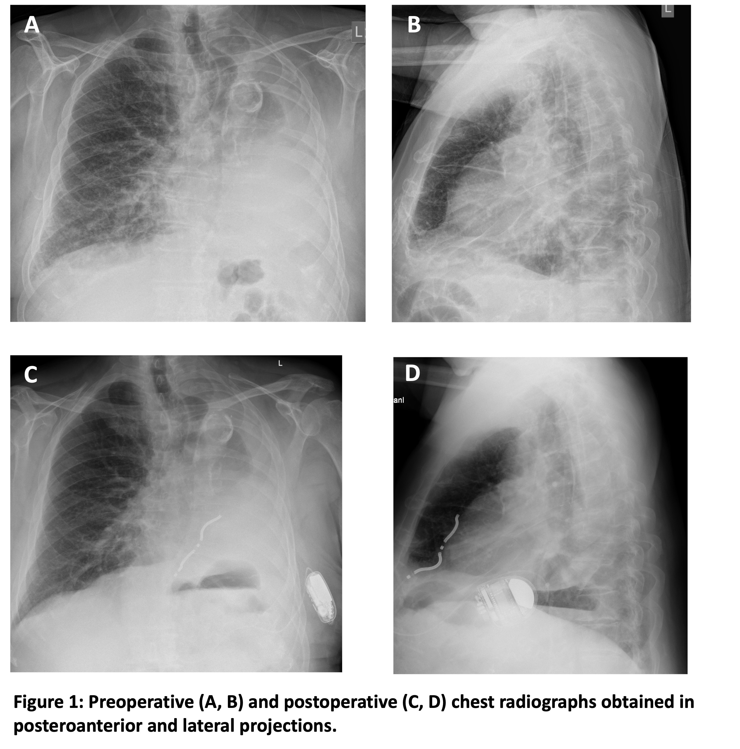
A 76-year-old man with ischemic cardiomyopathy and severely reduced left ventricular ejection fraction was referred for primary prevention ICD implantation. His medical history included advanced chronic kidney disease, severe mitral and tricuspid regurgitation, chronic obstructive pulmonary disease, peripheral arterial disease, and left-sided pneumonectomy for lung cancer 20 years earlier. Preprocedural imaging revealed marked mediastinal shift and cardiac rotation into the left hemithorax (Figure 1, A and B).
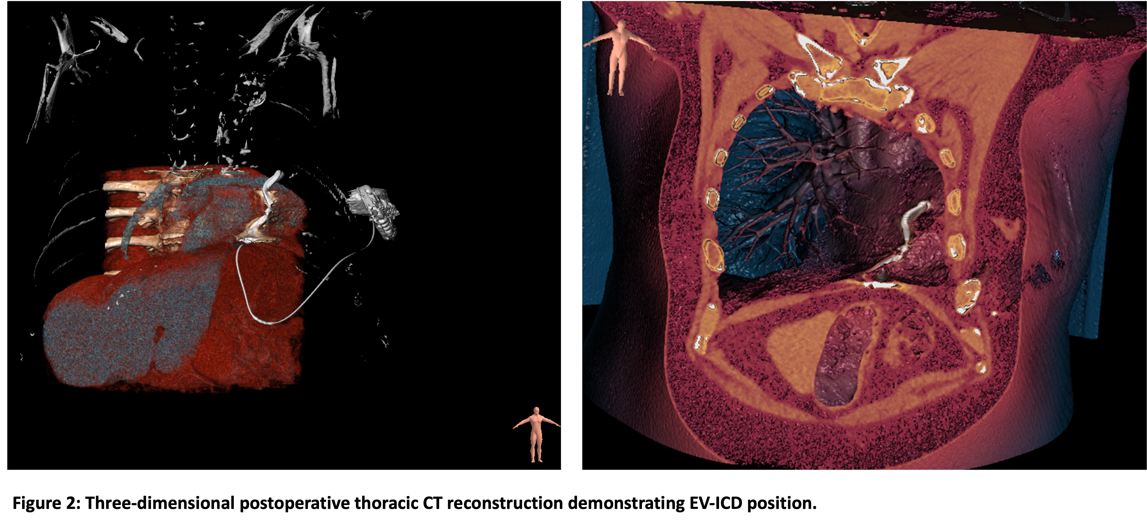
EV-ICD implantation was performed under deep sedation with propofol and ketamine and supported by non-invasive ventilation. Due to the altered anatomy, careful fluoroscopy-guided planning and substernal lead tunneling were required. A second tunneling attempt achieved optimal lead positioning. Final sensing parameters showed an R-wave amplitude of 1.2 mV and P-wave amplitude <0.2 mV. Defibrillation testing during implantation was not feasible because ventricular fibrillation could not be induced. Postoperative imaging, including chest radiography and computed tomography (CT) of the thorax, confirmed appropriate device and lead positioning (Figure 1, C and D and Figure 2). On the following day, successful defibrillation testing demonstrated termination of induced ventricular fibrillation with a 30-J shock, providing a 10-J safety margin. Device interrogation confirmed stable sensing and impedance values. The patient was discharged the next day without complications.
Discussion and Conclusion
This case demonstrates the feasibility and safety of EV-ICD implantation in a patient with highly complex thoracic anatomy after left-sided pneumonectomy. Deep sedation combined with non-invasive ventilation enabled successful implantation without endotracheal intubation. Careful procedural planning and fluoroscopic guidance were essential for successful substernal lead placement. This report expands the growing evidence supporting EV-ICD use in selected high-risk patients with challenging anatomical conditions.