Background:
Left ventricular free wall rupture is a rare but life-threatening complication of acute myocardial infarction. We report the clinical course of a patient with initial and recurrent hemorrhagic pericardial effusion and delayed myocardial rupture after progressive aneurysm formation.
Case Presentation: A 58-year-old man was admitted to our institution with an acute inferior ST-segment elevation myocardial infarction. Symptom onset had occurred the previous day. On admission, the patient presented in cardiogenic shock with severe metabolic acidosis (pH 7.1) and elevated serum lactate levels (7 mmol/L).
Initial transthoracic echocardiography demonstrated a severely reduced left ventricular systolic function with an ejection fraction of approximately 20% and a predominantly inferiorly located echogenic pericardial effusion (Fig. 1a). Computed tomography angiography excluded acute aortic dissection and classified the pericardial effusion as hemorrhagic. Subsequent emergency coronary angiography revealed severe three-vessel coronary artery disease. Successful recanalization of the occluded distal right coronary artery was performed.
Management and Clinical Course:
The patient required immediate catecholamine support and levosimendan treatment in the intensive care unit. Endotracheal intubation and mechanical ventilation were initiated immediately. Due to multiorgan failure, broad-spectrum antibiotic therapy and temporary renal replacement therapy were required.
After five days, the patient was successfully extubated. During further recovery, two electrical cardioversions were performed for atrial fibrillation. Follow-up echocardiography demonstrated transformation of the initially echogenic pericardial effusion into an echo-free effusion accompanied by progressive thinning of the inferoposterior left ventricular wall (Fig. 1b).
After three weeks, the patient had clinically stabilized and was scheduled for cardiac surgery. However, despite detailed counseling regarding the associated risks, he left the hospital against medical advice despite recommendations for surgical treatment.
Seven weeks after the initial infarction he was readmitted following a syncopal episode. Echocardiography now demonstrated a large inferoposterior left ventricular aneurysm with a recurrent hemorrhagic pericardial effusion (Fig. 1c). Emergency surgery was performed. Intraoperatively, fresh intrapericardial hemorrhage and an inferoposterior ventricular aneurysm with active oozing were identified (Fig. 1d). Surgical repair was successfully accomplished using the Dor procedure (Fig. 1 e, f). The postoperative course was uneventful.
Conclusion: Retrospective analysis of serial imaging suggested that the initial hemorrhagic pericardial effusion represented a contained slit-like left ventricular free wall rupture (Fig. 1, arrows). Subsequent imaging documented progressive wall thinning, liquefaction of the pericardial hematoma, and gradual aneurysm formation. Seven weeks after the index infarction, this process culminated in a large inferoposterior aneurysm with a recurrent hemopericardium (Fig. 1c, star) caused by delayed free wall rupture. This case illustrates the dynamic evolution of a contained ventricular free wall rupture and highlights the importance of serial imaging and timely surgical intervention.
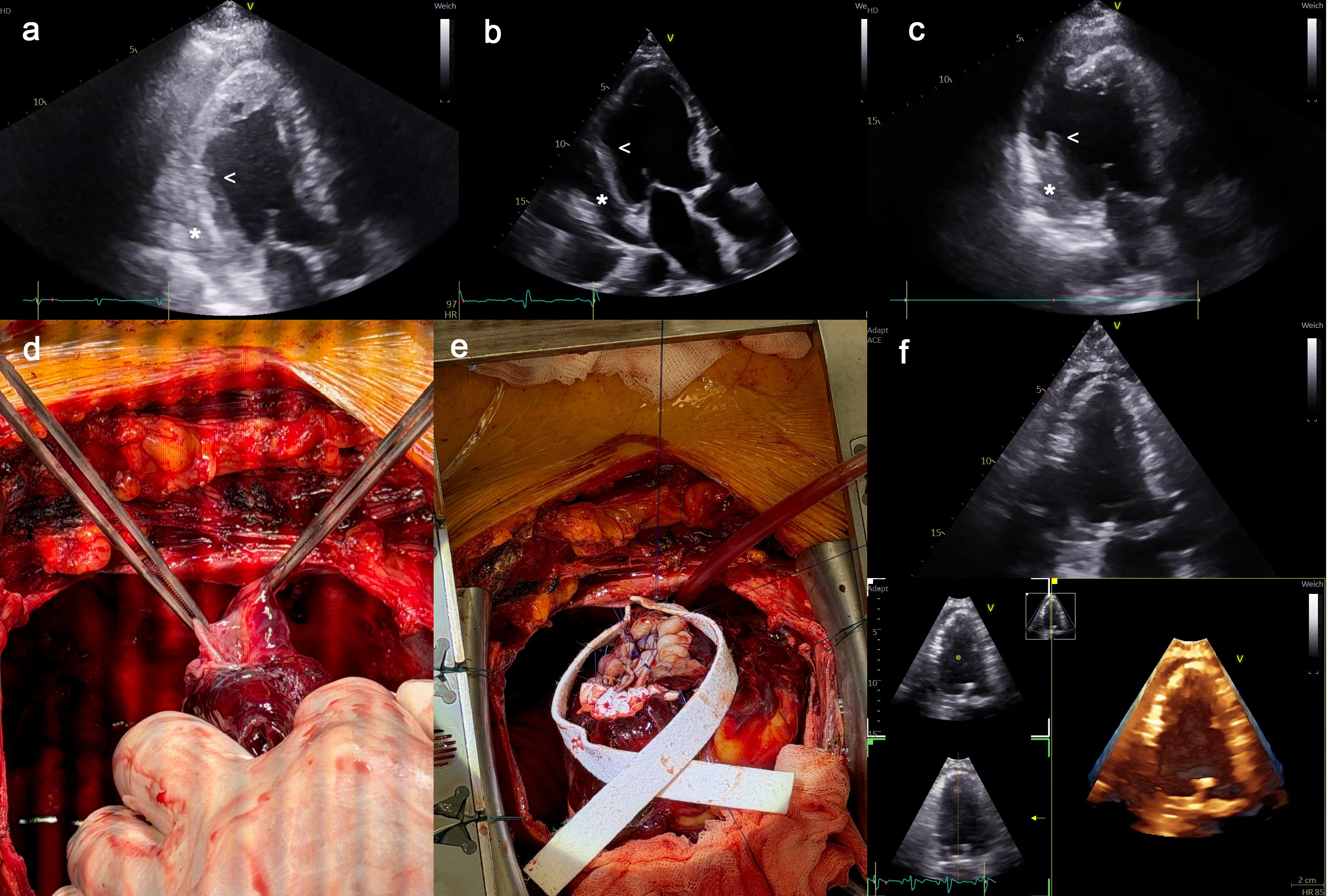
Figure 1: Contained left ventricular free wall rupture progressing to inferoposterior aneurysm formation and delayed rupture with surgical repair



