Background
Retinal vessel analysis is a non-invasive tool to assess microvascular function. Retinal venular widening in static analysis and an impaired flicker light induced dilation (FID) of retinal vessels in dynamic analysis is known to be associated with higher cardiovascular risk. The course of endothelial function assessed by retinal vessel analysis in patients with acute myocardial infarction and during follow-up optimizing cardiovascular risk factors, especially optimal lipid-lowering therapy is so far unknown. Therefore, the aim of the present study was to investigate retinal vessel analysis in patients during the hospital stay of the index event and during long term follow-up on optimal lipid-lowering therapy.
Methods
We performed static retinal vessel analysis (SVA) and dynamic retinal vessel analysis (DVA 3.0) applying flicker light stimuli in 15 patients post STEMI (median within 3 days) and 5 years after the index event. At baseline all patients received a combination of high dose statin therapy (atorvastatin 80 mg) and ezetimibe. We obtained cardiovascular risk status at baseline and during follow-up including smoking status, diabetes mellitus, blood pressure, lipid lowering therapy and lipid profile. One patient was excluded due to relevant retinal abnormalities with influence on retinal vessels. In two patients the quality of the measurements was insufficient. We included 12 patients in the final analysis.
Results
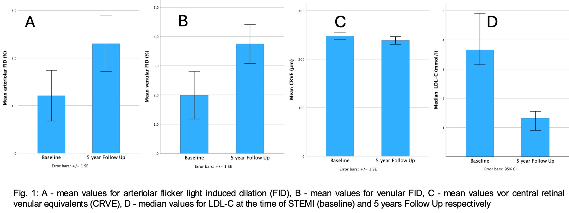
Five years post STEMI all except for two patients still were treated with a combination of statin and ezetimibe. Two patients were on triple lipid lowering with a high intensity statin, ezetimibe and PCSK9 inhibitor. Most patients had a previous diagnosis of arterial hypertension, but presented with well-controlled blood pressure (mean 128 ± 4.5 mmHg) at 5 year follow-up. Out of 9 smokers at the time of STEMI 6 had given up until follow-up. The majority of patients reached an LDL cholesterol <1.4 mmol/l for the high-risk group according to the current ESC guidelines for dyslipidemias (LDL-C at baseline median 3.66 mmol/l IQR 1.56 mmol/l, LDL-C at follow-up median 1.32 mmol/l IQR 0.58 mmol/l).
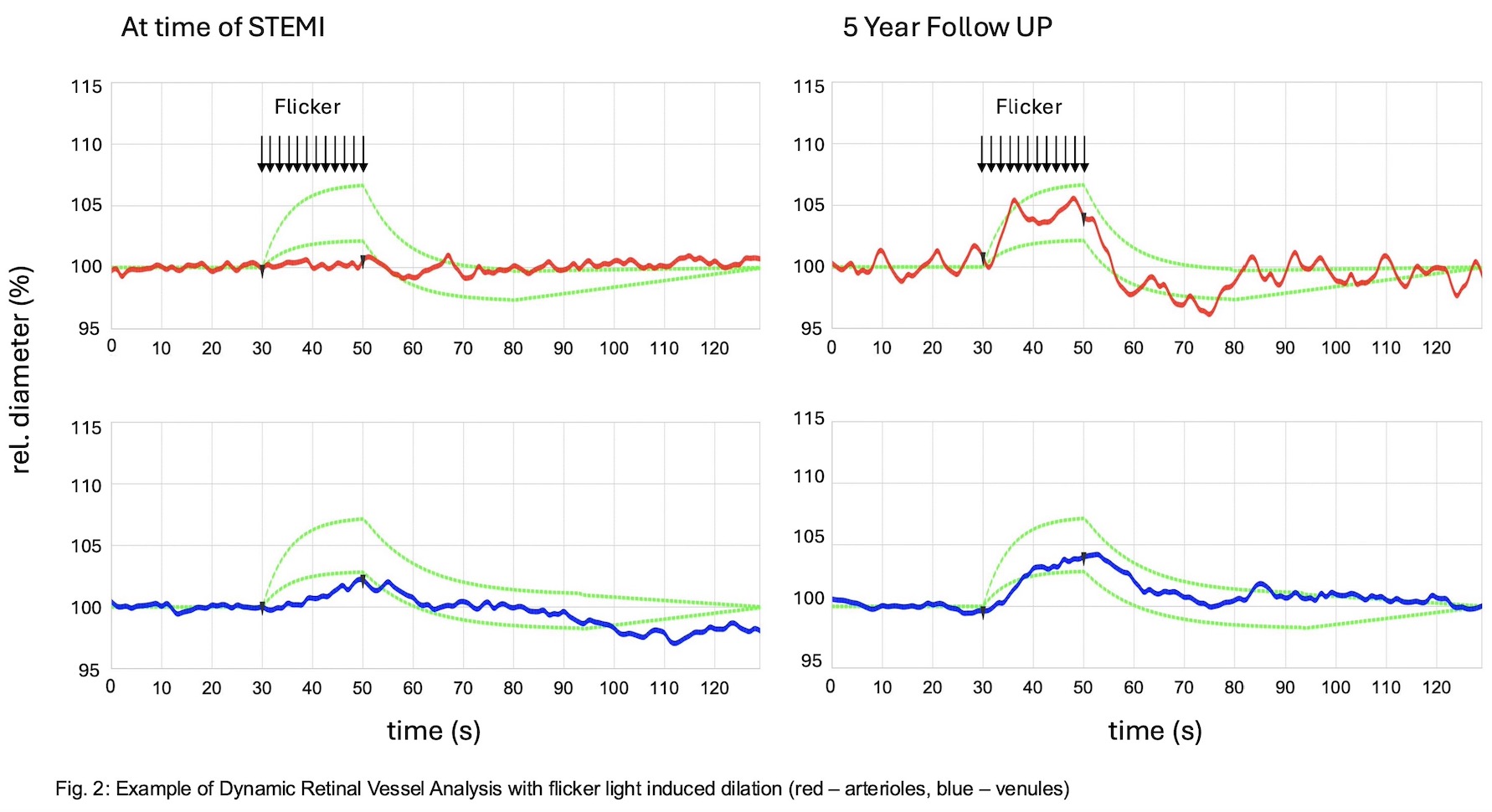
As expected, static retinal vessel analysis (SVA) revealed lower values for central retinal venular equivalents (CRVE) (one-sided p < 0.036) during follow-up compared to baseline. Dynamic retinal vessel analysis (DVA 3.0) showed higher values for FID of retinal arteries (one-sided p = 0.027) and veins (one-sided p = 0.043) during follow-up (see figure 1 and figure 2).
Conclusion
In patients after ST-elevation myocardial infarction with optimized cardiovascular risk factors, especially intensive lipid-lowering therapy reduces LDL-C and is associated with improved microvascular dysfunction assessed by retinal vessel analysis during long term follow-up. Further studies are needed to assess retinal vessel analysis as a tool to improve cardiovascular risk prediction in high risk patients.