Background:Pulsed-Field Ablation (PFA) is increasingly being used for catheter ablation of ventricular tachycardia (VT), either alone or in combination with radiofrequency(RF) ablation. Data on the acute feasibility and safety of the focal dual-energy TactiFlex Duo catheter (Abbott) are not yet available. This study reports the first clinical experience with VT ablation using the TactiFlex Duo catheter.
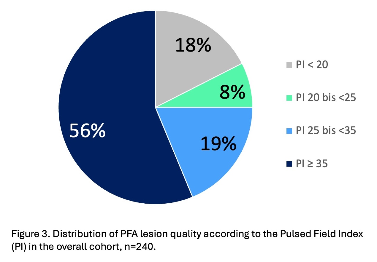
Methods:All consecutive patients undergoing VT ablation with the TactiFlex Duo catheter between 04/2026 and 06/2026, were prospective enrolled. Clinical, procedural, and acute outcome data, as well as PFA-specific metrics, including Pulsed Field Index (PI) lesions were assessed. According to the manufacturer's definition, lesion quality was classified as highest quality (PI ≥ 35), high quality (PI 25-34), good quality (PI 20-24), and low quality (PI < 20).
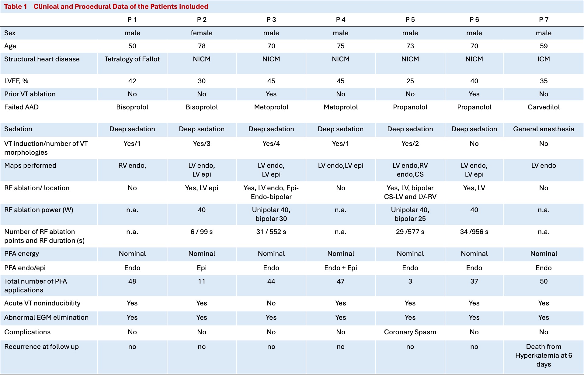
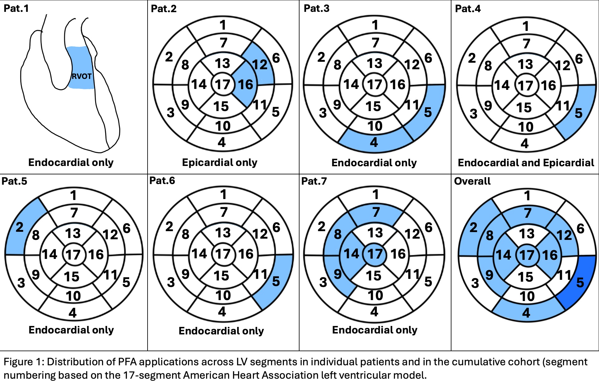
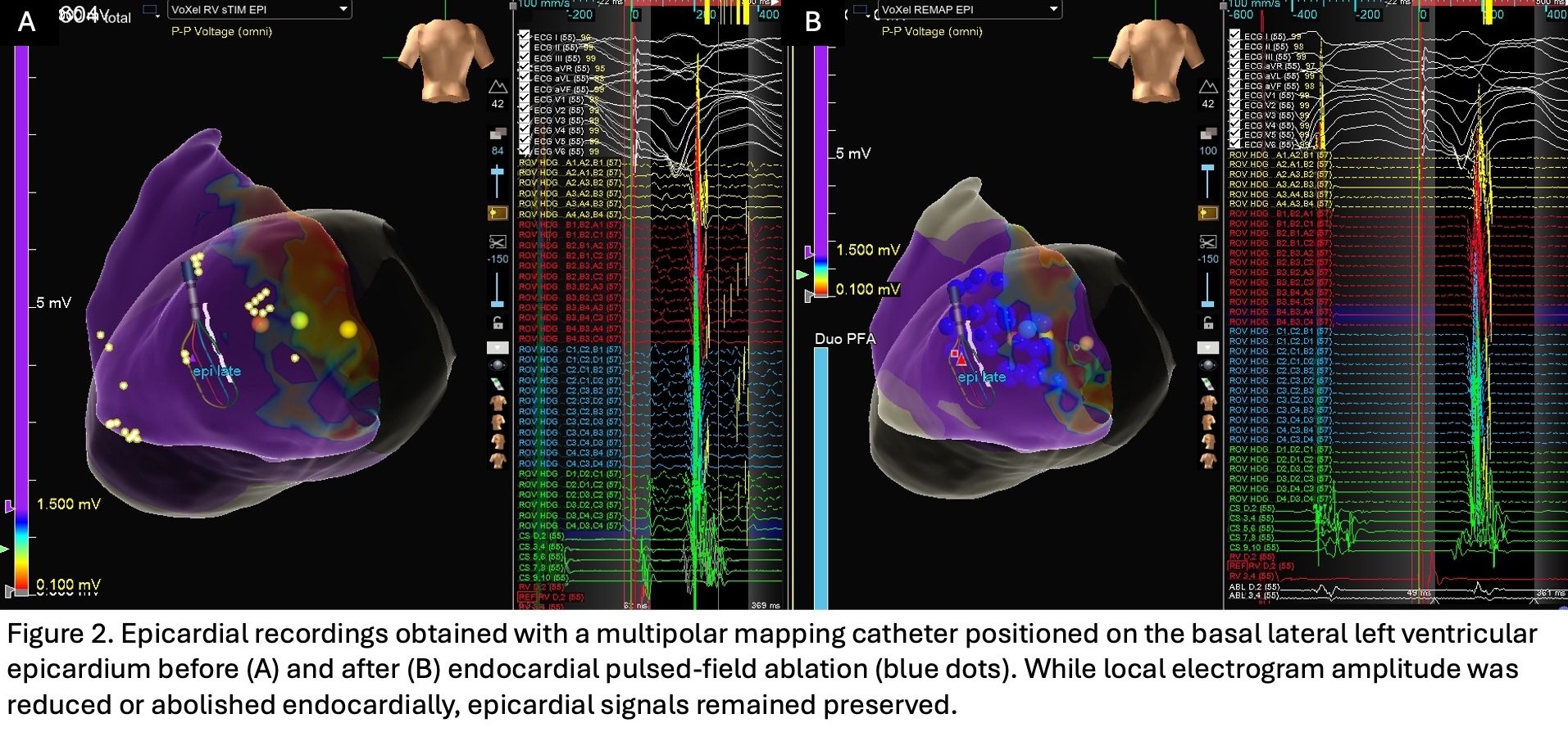
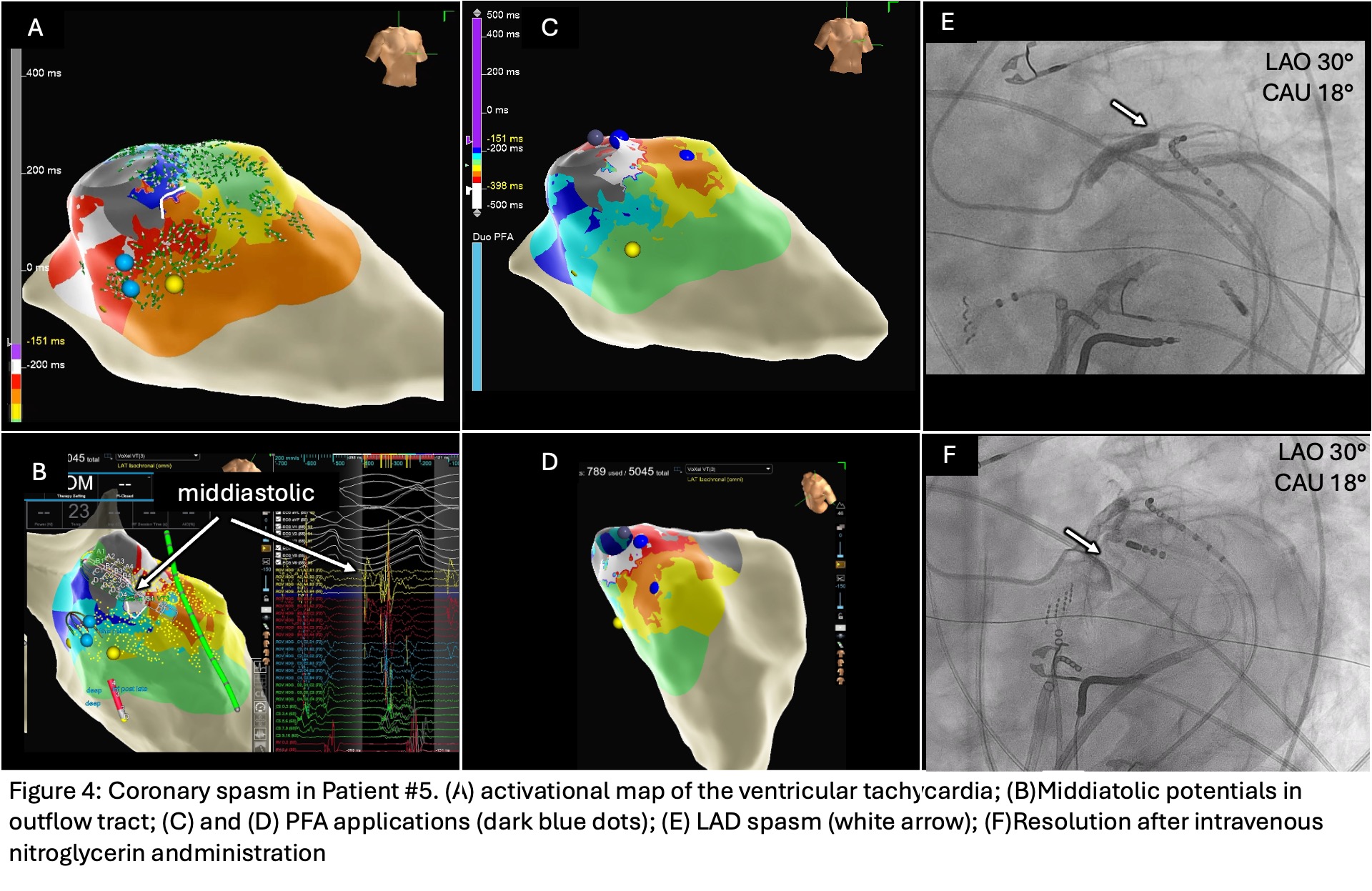
Results:A total of 7 patients (6 male; median age 70 years; median LVEF 40%)were included. Clinical and periprocedural characteristics of all patients are presented in Table 1. Endocardial mapping was performed in all patients and in 4 patients additionally epicardial. PFA was delivered using nominal energy with a target of 5 pulses per application, in 5 patients endocardially only (one RVOT, n=1; LV, n=4), both endocardially and epicardially in one patient, and epicardially only in one patient (Fig. 1). To assess the transmurality of PFA, endocardial PFA applications were delivered to the basal lateral LV segment in 3 patients, with an HD‑Grid catheter positioned on the directly opposing epicardial surface. In a 4th patient, endocardial PFA was performed in the LVOT while a decapolar catheter was in the great cardiac vein. In both settings, endocardial and epicardial electrograms were monitored throughout energy delivery to capture the dynamics of EGM change. In all 4 cases, a significant reduction or disappearance of the local EGM amplitude was observed endocardially, but not epicardially (Fig.2). Additional RF ablation, including bipolar ablation in 2 patients, was performed in 4 patients with a median RF duration of 564 s (IQR 439-672). A total of 240 PFA lesions were delivered, with a median of 44 lesions per patient (IQR 24-48). Of all PFA lesions 56% were classified as highest quality (PI ≥35), 19% as high quality, 8% as good quality and 18% as low quality (Fig. 3). Per patient a median of 45% of lesions (IQR 31-80) were of highest quality. During PFA delivery median average contact force was 11,6g (IQR 10-19,8). Non-inducibility of any VT was achieved in 6 patients. One patient developed a spasm of the left anterior descending coronary artery after two PFA applications delivered endocardially in proximity to the vessel, resolving after intravenous nitroglycerin administration (Fig. 4). No other periprocedural complications, including ICD malfunction, were observed. During 1 month follow-up six patients remained VT free. One patient with VT inducibility at the end of ablation procedure subsequently underwent stereotactic arrhythmia radioablation. One patient died 9 days after ablation due to severe hyperkalaemia leading to pulseless electrical activity.
Conclusion:In this first reported case series,VT ablation using TactiFlex Duo catheter appeared feasible and effective for acute outcomes in patients with structural heart disease. Most PFA lesions were of high quality. During endocardial PFA near coronary vessels coronary spasm may occur.