BackgroundThe right ventricle (RV) differs fundamentally from the left ventricle (LV) in terms of embryological origin, anatomy, loading conditions and physiological function, yet remains underrepresented in cardiovascular research. Human models to investigate RV biology are scarce, and current experimental approaches predominantly focus on the LV. Therefore, this study aimed to establish and functionally characterize human RV living myocardial slices (LMS) and identify determinants associated with RV-LMS viability during electromechanical culture.
MethodsHuman RV myocardium was obtained from explanted failing hearts of patients undergoing heart transplantation. Precision-cut myocardial slices were generated using a vibratome and cultured in biomimetic chambers under continuous electromechanical stimulation at 1 Hz, assessing contraction amplitude, diastolic tension, time-to-peak (TTP), and relaxation time 80% (RT80). Slice viability was monitored and associations with donor characteristics, myocardial fibrosis, and chamber resistance were analyzed.
ResultsRV-LMS were generated from 15 explanted failing hearts (34 slices) and maintained stable contractile activity during 8 days of electromechanical culture. Contractile amplitude showed a non-significant increase during the first days of culture and remained stable thereafter, while diastolic tension was preserved (Fig.1B). Contraction kinetics changed over time, with a shortening of TTP (Fig.1C) and a trend towards shorter RT80 (Fig.1D), suggesting an early adaptation phase. Masson Trichrome (MT) staining and histological quantification revealed significantly higher fibrosis levels in slices with loss of viability than in viable preparations (21.6% vs. 11.3%, p<0.0001) (Fig.1E-F). Most RV-LMS remained viable, however, 7 slices from 3 hearts developed contractile insufficiency and subsequently lost viability (Fig.1H).
Among all baseline characteristics, donor BMI was the only parameter associated with loss of RV-LMS viability in logistic regression analysis (OR 2.81, 95% CI 1.26–13.77, p=0.007), with higher BMI values observed in donors yielding non-viable slices (27.3±2.1 vs. 23.4±1.9 kg/m², p=0.008). Finally, while chamber resistance did not affect functional properties of surviving RV-LMS, slices cultured under normal-resistance conditions more frequently developed contractile insufficiency and loss of viability compared with low-resistance chambers (p=0.01 and p<0.01, respectively).
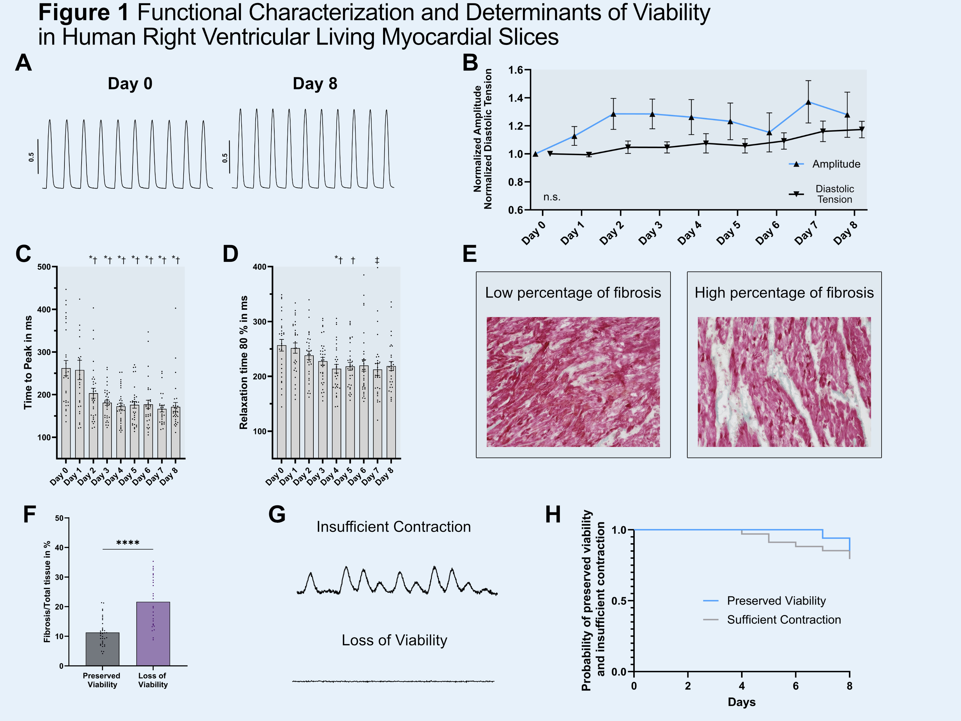 Figure 1. (A) Original twitch traces day 0 and 8. (B) Normalized amplitude, diastolic tension, (C) TTP and (D) RT80; symbols indicate intra-group comparisons (*p<0.01 vs. day 0; †p<0.05 vs. day 1; ‡p<0.05 vs. day 0). (E) Representative MT stained LMS and (F) comparison of fibrosis level in LMS with preserved/lost viability;(****p<0.0001). (G) Representative LMS with insufficient contraction/lost viability. (H) Kaplan-Meier analysis of LMS with sufficient contraction and preserved viability. Data as mean±SEM.Conclusion
This study provides the first systematic functional characterization of human RV-LMS during electromechanical culture. RV-LMS maintained stable contractile activity over several days and emerged as a feasible and translationally relevant platform for studying human RV biology. Importantly, myocardial fibrosis, donor BMI and culture conditions influenced slice viability, highlighting intrinsic and extrinsic factors that should be considered in future RV-LMS studies and therapeutic applications.



