Cardiovascular remodeling is a hallmark of myocardial infarction, hypertension, and heart failure, characterized by fibrotic changes and impaired Ca²⁺ homeostasis. Excessive fibroblast activation and extracellular matrix deposition increase myocardial stiffness and drive dysfunction, while disrupted intracellular Ca²⁺ cycling impairs excitation–contraction coupling, contractility, and electrical stability. Ca²⁺ signaling is tightly regulated by plasma membrane ion channels, the sarcoplasmic reticulum (SR), and mitochondria, which coordinate Ca²⁺ release, reuptake, buffering, and metabolic coupling. MicroRNAs (miRNAs) have emerged as key post-transcriptional regulators of these processes. In particular, miR-16-5p, miR-125b-5p, and let-7a-5p regulate fibrotic and Ca²⁺-handling pathways, suggesting potential roles in cardiac remodeling.
This study investigated whether combined inhibition of miR-16-5p, miR-125b-5p, and let-7a-5p alters fibrotic signaling and Ca²⁺-handling pathways in vivo. Fifteen-week-old female C57Bl/6J mice received intraperitoneal antagomirs every 3 days for 14 days (10 mg/kg each; total 30 mg/kg). Cardiac tissue was analyzed by histology and western blotting for fibrosis markers and Ca²⁺-handling proteins spanning sarcolemmal (CaV1.2, NCX1), SR (RyR2, SERCA2, CASQ, PLN), and mitochondrial (MCU complex components and regulators) systems.
Histological staining (Masson’s Trichrome, Sirius Red) showed no overt collagen deposition in antagomir-treated hearts. However, protein analysis revealed increased collagen type I (COL1) and SMAD3, alongside reduced TGF-β1, indicating early activation of pro-fibrotic signaling without structural fibrosis.
Analysis of Ca²⁺-handling proteins demonstrated broad remodeling across compartments. Membrane-associated CaV1.2 and NCX1 were significantly downregulated. Within the SR, RyR2 expression increased, SERCA2 remained unchanged, and both CASQ and PLN were reduced, indicating disrupted Ca²⁺ storage and regulation with a shift toward enhanced release capacity. Mitochondrial Ca²⁺ regulatory proteins MCUR1, MICU1, and MICU2 were decreased, while MCU remained stable. Regulatory proteins UCP2, UCP3, and PRMT1 were also reduced, suggesting impaired mitochondrial Ca²⁺ buffering and altered metabolic coupling.
Overall, combined inhibition of miR-16-5p, miR-125b-5p, and let-7a-5p induced coordinated molecular changes in fibrotic signaling and multi-compartment Ca²⁺ handling prior to detectable structural fibrosis. These findings suggest that these miRNAs contribute to maintenance of Ca²⁺ homeostasis and suppression of early pro-fibrotic signaling in the heart, and their inhibition triggers a pre-fibrotic, pro-remodeling molecular state.
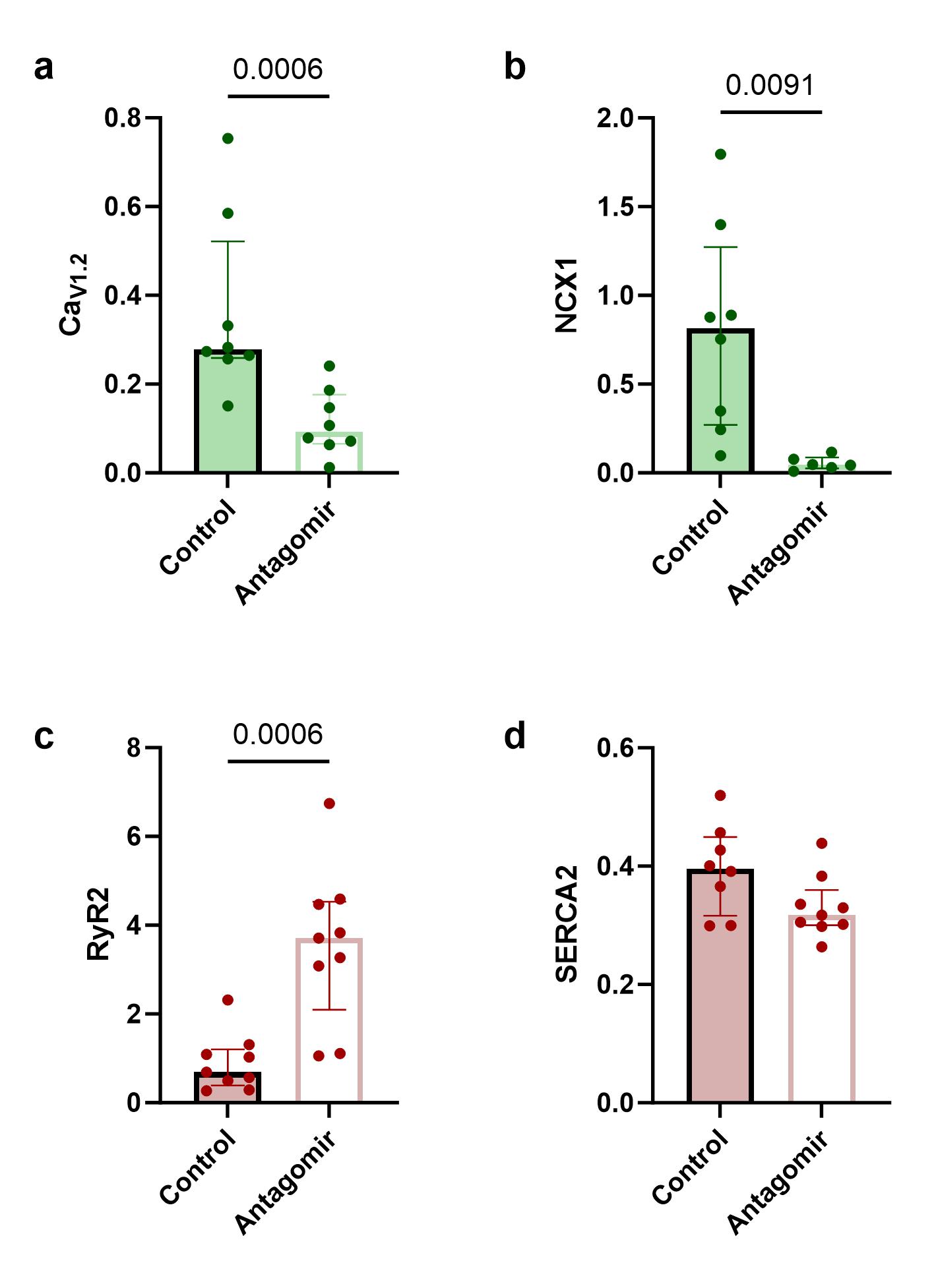
Figure 1: Quantification of membrane-bound (a) CaV1.2, (b) NCX1 as well as SR-bound (c) RyR2 and (d) SERCA2 in cardiac tissue of control and antagomir treated mice. Bars are given as median with IQR. CaV1.2: voltage-dependent L-type calcium channel; NCX1: sodium-calcium exchanger 1; RyR2: ryanodine receptor 2; SERCA2: sarcoplasmic/endoplasmic reticulum calcium ATPase 2
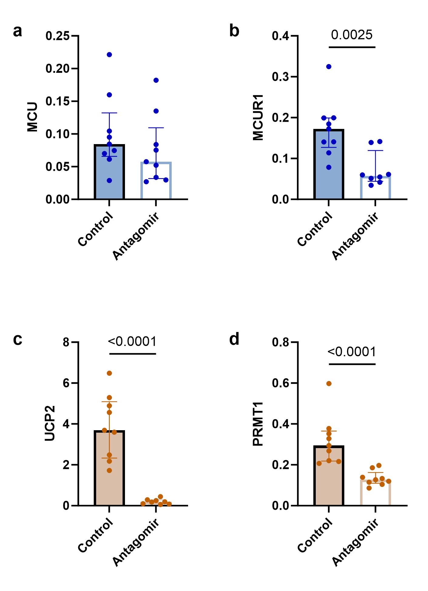
Figure 2: Quantification of mitochondrial (a) MCU, (b) MCUR1, (c) UCP2, and (d) PRMT1 in cardiac tissue of control and antagomir treated mice. Bars are given as median with IQR. MCU: mitochondrial calcium uniporter; MCUR1: mitochondrial calcium uniporter regulator 1; PRMT1: protein arginine N-methyltransferase 1; UCP2: uncoupling protein 2



