Background
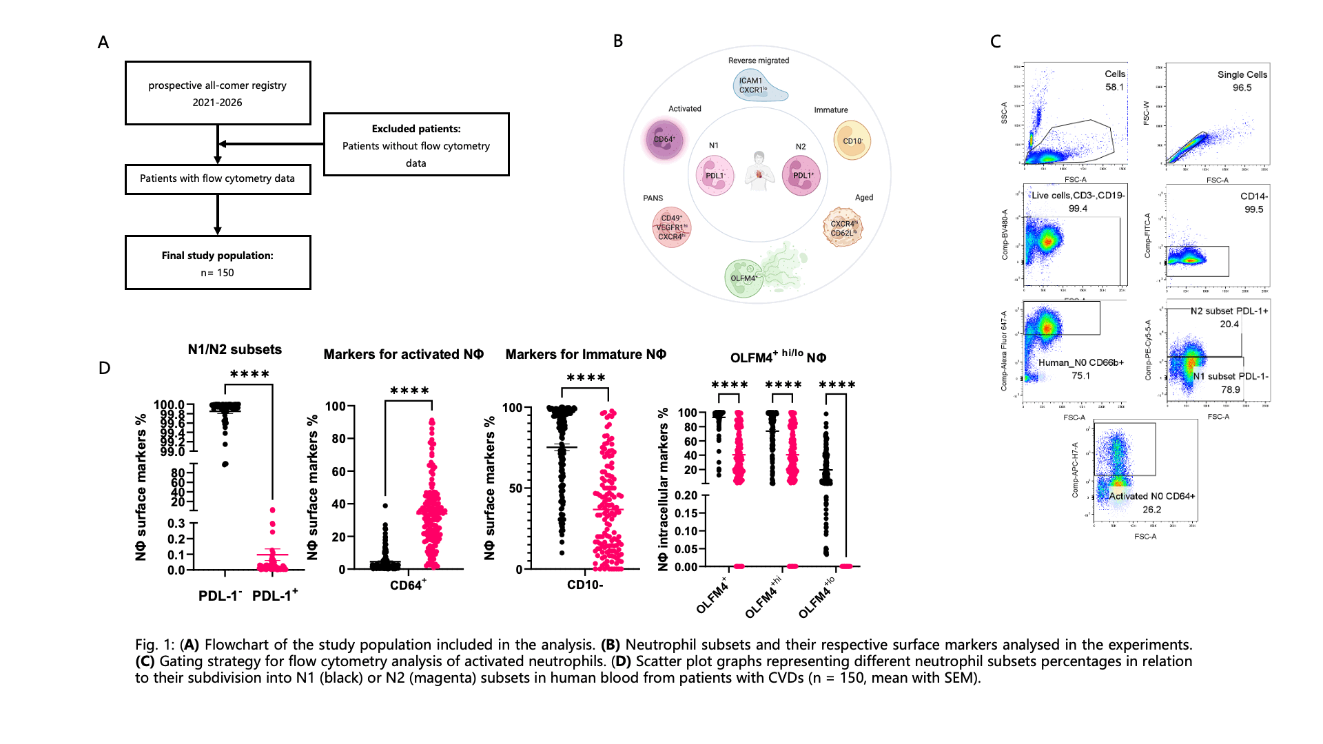
Neutrophils (NΦ) are the first leukocytes to arrive at cardiac injury sites. They aid in both tissue damage and repair as neutrophils divided into pro-inflammatory (N1) and anti-inflammatory (N2) subsets. Several neutrophil subsets need to be explored in cardiovascular diseases, that have been identified in other disease models. Neutrophils also exhibit plasticity and hence characterizing the following subsets will help to understand the mechanism by which these subsets contribute to tissue inflammation or repair that help in the future to develop targeted therapies. They can firstly be divided into N1 (PDL-1-) versus N2 (PDL-1+) neutrophils and then be further divided into activated (CD64+), pro-angiogenic ((PANs) CD49d+, VEGFR1hi, CXCR4hi), aged (CXCR4hi, CD62Llo), immature (CD10-) and reverse-migrated neutrophils (ICAM1hi, CXCR1lo). Moreover, they can be analysed for the intracellular marker OLFM4, high expression of which suggests an enhanced capacity to perform NETosis.
Methods & Result
A total of 150 patients (mean age 66 years (SD): ± 14 years); 46.3% ♀) from a prospective all-comer registry were analysed. Neutrophils were isolated from peripheral blood samples, followed by quantitative analysis of neutrophil subset markers via FACSymphony A1. Majority of neutrophils showed no expression of the marker PDL-1, specifying them as N1 neutrophils. Further staining suggested that neutrophils cannot be unequivocally categorized into distinct subsets but rather overlap across multiple subsets. Therefore N1 (PDL-1-) as well as N2 (PDL-1+) neutrophils can be further subdivided. The analysis showed that N2 neutrophils presented a broader range of surface markers than N1 neutrophils within the activated (CD64+), the pro-angiogenic ((PANs) CD49d+, VEGFR1hi, CXCR4hi), the aged (CXCR4hi, CD62Llo) and the reverse-migrated neutrophils (ICAM1hi, CXCR1lo). Consequently, N2 neutrophils showed greater overlap across subsets, whereas N1 neutrophils grouped into more distinct subsets. Meanwhile, N1 neutrophils showed a decreased expression of CD10, a marker associated with neutrophil maturation. Furthermore, N1 neutrophils exhibited increased intracellular OLFM4 expression, with a particular increase in the high-expression population.
Conclusion
Neutrophils may represent a transitional or mixed subset rather than strictly defined lineage. Collectively the data indicate that N1 neutrophils are a rather immature and plastic population, while N2 neutrophils are more mature and have undergone several differentiation processes. Consistently, N1 neutrophils are part of the high-OLFM4-expressing population, potentially contributing to enhanced pathogen defence as the so-called pro-inflammatory subset, whereas N2 neutrophils show lower expression levels of OLFM4 thus having lost their NETosis capacity, accordingly to their supposed anti-inflammatory character. Going forward, identification of patient specific neutrophil phenotypes in correlation with clinical parameters will be conducted, which could reveal new therapeutic targets and thereby enable reduction of cardiac damage.