Background/Purpose:
Monocyte-endothelial interactions drive acute inflammation and chronic vascular disease in type 2 diabetes mellitus (T2DM). However, it remains unclear whether molecular pathways involved in TNF-α-induced and diabetes-associated monocyte trafficking are similar or divergent. Therefore, the aim of this study is to explore potential divergences.
Methods:
We utilized a physiological flow-based imaging system under controlled shear stress to quantifyCD14⁺ monocyte adhesion, transendothelial migration (TEM), and abluminal residence on human umbilical vein endothelial cell (HUVEC) monolayers after activation of those cells by either TNF- α or serum obtained from T2DM patients.
Results:
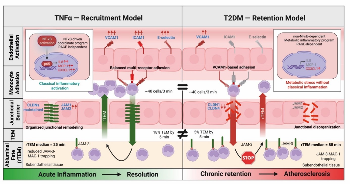
Both stimuli resulted in similar initial monocyte adhesion (40 adherent cells by 3 minutes within view field). On the other hand, TEM efficiencies were quite variable. TNF-α stimulation promoted TEM (18% by 5 min post initiation) whereas T2DM conditions showed severely impaired TEM (~5% at 5 min post initiation).
Moreover, monocytes under T2DM conditions were retained abluminally longer than TNF- α (median 85 min vs. 25 min; p < 0.0001). This prolonged retention phenotype was mimicked by exposing endothelial cells to hyperglycemia.
To elucidate the underlying molecular mechanisms, we performed a comparative RNA-sequencing analysis of T2DM-stimulated (GSE92724) and TNF- α stimulated (GSE134489) endothelial cells. Analysis revealed a near-zero transcriptional correlation (r = 0.018), indicating highly divergent genomic responses. TNF- α stimulation resulted in a coordinate, NF-KB–dependent upregulation of VCAM1, ICAM1, E-selectin, and key junctional molecules, leading to organized junctional remodeling. Conversely, T2DM conditions led to a VCAM1-biased adhesion pattern with ICAM1 downregulation (0.61-fold), claudin suppression, and junctional disorganization.
KEGG pathway mapping further confirmed that TNF- α promotes coordinated endothelial–leukocyte integrin co-activation, whereas T2DM causes discordant junctional loss. Subsequent qPCR validation showed selective upregulation of RAGE and JAM-3 under T2DM conditions—consistent with AGE-RAGE signaling and JAM-3–MAC-1-mediated monocyte trapping—in the absence of classical NF-KB activation.
Conclusion:
We could show different patterns of monocytic adhesion and TEM in T2DM compared to TNF-α. TNF-α stimulates leukocyte recruitment while T2DM induces a leukocytic retention by VCAM-1 biased adhesion, junctional disorganization and impaired reverse transendothelial migration. The present study identifies the AGE-RAGE axis and JAM-3–MAC-1 interactions as distinct, promising therapeutic targets for mitigation of diabetic vascular inflammation.