IntroductionCardiovascular disease remains the leading cause of death worldwide. Especially ischemia/reperfusion injury (IR) after myocardial infarction is associated with high mortality due to malignant arrhythmias and heart failure. The Leukemia Inhibitory Factor (LIF) has emerged as a cytokine with potential cytoprotective properties, including modulation of cellular responses to oxidative stress. Because oxidative stress is a major driver of cardiomyocyte injury during IR, we hypothesized that LIF may exert cardioprotective effects under IR conditions.
MethodsMurine ventricular cardiomyocytes were isolated via retrograde perfusion of explanted wildtype mouse hearts on a Langendorff perfusion apparatus. IR was modelled
in vitro by 75 min hypoxia (in Esumi buffer) and 14 h of subsequent reoxygenation. Human induced pluripotent stem cells (iPSCs) were differentiated into cardiomyocytes using a WNT signalling activation/inhibition protocol followed by a maturation phase until day 30. In human iPSC-cardiomyocytes, IR was modelled by 120 min hypoxia (in Esumi buffer) and 14 h of subsequent reoxygenation. During reoxygenation, cardiomyocytes were either left untreated (IR group) or treated with 10 ng/mL LIF (IR + LIF group). Electrically stimulated Ca
2+ transients were analysed in Fura-2 AM-loaded cardiomyocytes using epifluorescence microscopy. Action potentials were recorded using whole-cell current-clamp technique.
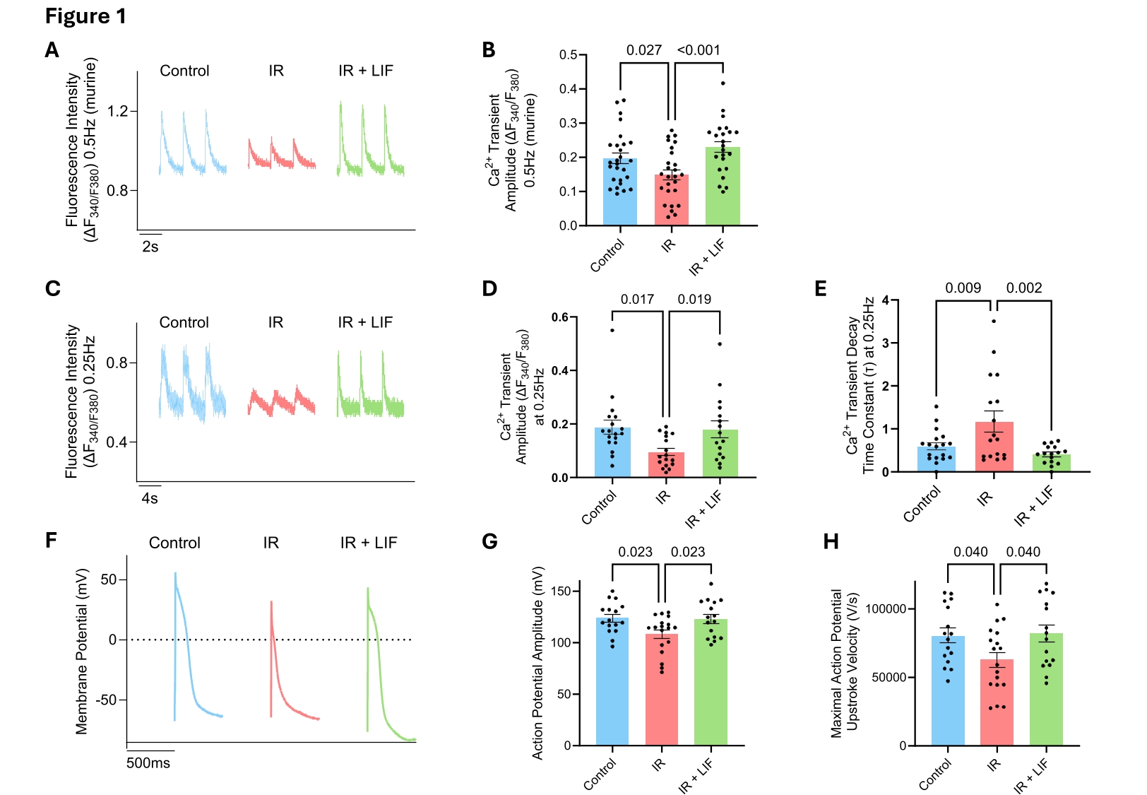
ResultsUpon IR, murine cardiomyocytes showed a decreased Ca
2+ transient amplitude from (in ΔF
340/F
380) 0.197 to 0.148 (Fig. 1A+B). Notably, LIF treatment completely protected from this decline and cardiomyocytes showed a Ca
2+ transient amplitude of 0.231 ΔF
340/F
380 (p<0.001 vs. IR; Fig. 1A+B). To enhance the translational impact of our study, we transferred this finding to human iPSC-derived cardiomyocytes.
Human iPSC-derived cardiomyocytes subjected to IR showed a significant decrease in Ca
2+ transient amplitude, falling to approximately 50% of control levels, from (in ΔF
340/F
380) 0.189 to 0.096 (at 0.25 Hz; Fig. 1C+D). Similar findings were observed at electrical stimulation rates of 0.5 Hz, 1 Hz, and 1.5 Hz (data not shown). The Ca
2+ transient decay time constant (τ) was significantly increased upon IR in non-treated human cardiomyocytes (Fig. 1E). LIF-treated cardiomyocytes were protected against these IR-induced aberrations of cellular Ca
2+ homeostasis (Fig. 1C-E).
In addition, untreated human iPSC-derived cardiomyocytes subjected to IR showed a significantly reduced action potential amplitude (from 124.0 mV to 108.3 mV), accompanied by a corresponding decrease in maximal action potential upstroke velocity (from 80748 V/s to 62815 V/s; Fig. 1F-H). LIF treatment during IR protected from these pathological electrical alterations and human iPSC-cardiomyocytes showed a significantly increased action potential amplitude and maximal action potential upstroke velocity (vs. IR without LIF; Fig. 1F-H).
ConclusionWe observed that the cytokine LIF confers cardioprotection by preserving Ca
2+ transient and action potential properties in an
in vitro model of ischemia/reperfusion injury. Ongoing work aims to elucidate the underlying molecular mechanisms and to further evaluate the protective potential of LIF
in vivo using a mouse model of surgically induced myocardial infarction.