Introduction:
Engineered heart tissues (EHTs) have emerged as a powerful system for cardiovascular disease modelling, drug testing and mechanistic studies of human cardiac physiology. As spontaneously beating human myocardial tissues, EHTs close the gap between cell culture systems and ex vivo models, allowing the study of cardiac function in a physiologically relevant human tissue context. Contractile properties and some electrophysiological features of cardiomyocytes in EHT context, such as action potentials, are well defined. Other critical electrophysiological properties of EHT however, like conduction velocity (CV) at baseline, are still to be fully established at tissue level, in order to use EHT as a meaningful tool to study arrhythmogenesis. Optical mapping approaches capable of quantifying these parameters in EHTs could substantially expand their utility for disease modelling and preclinical pharmacological testing.
Aim:
We aimed to establish a voltage-sensitive optical mapping platform for EHTs that enables high-resolution assessment of action potential propagation and restitution properties during unilateral pacing.
Methods:
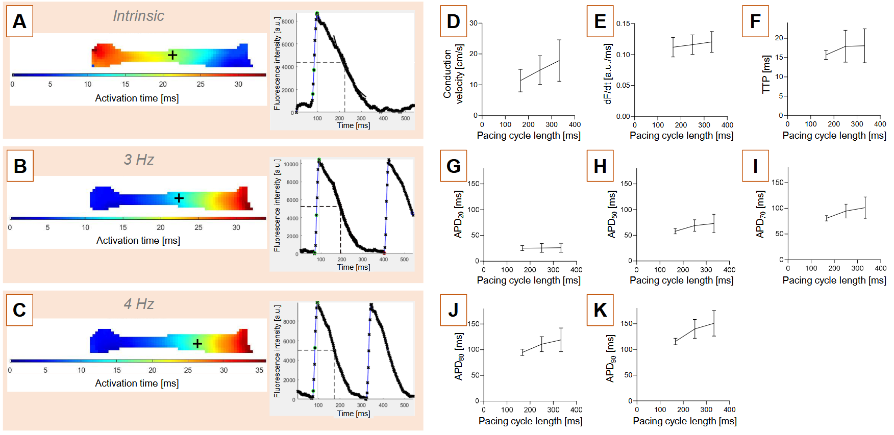
Optical mapping in EHT was carried out using a custom-built setup, comprised of a gas and temperature-controlled bath and a high speed back-illuminated Camera. Image acquisition and analysis, were performed using open source/custom built software. Dye loading was performed by individually incubating EHTs with a red-shifted voltage sensitive dye (RH 237). Pacing threshold was determined for each EHT individually and double threshold was used for pacing. To investigate impulse propagation under controlled conditions, optical mapping was combined with an asymmetric field stimulation protocol enabling unilateral excitation of EHTs: Asymmetrical electrical pacing units were placed at the left post of the EHT in order to avoid simultaneous activation of the whole EHT and pacing was carried out at 3, 4, and 6 Hz for atrial and 2 and 3 Hz for ventricular EHTs. EHTs were electromechanically uncoupled with 11 µM of blebbistatin in the superfusion chamber.
Results:
Using an optical mapping setup adapted for EHT recordings, we obtained high-resolution voltage signals across the surface of intact EHTs. Analysis of spontaneous activity revealed the presence of distinct intrinsic pacemaker regions within individual EHTs. Activation consistently originated from areas adjacent to one of the two posts. Intrinsic beating frequencies determined from bright-field recordings closely matched cycle lengths (CL) derived from voltage-sensitive dye recordings, demonstrating concordance between mechanically (574 ms ±22 CL) and electrophysiologically (558 ms ± 52 CL) assessed spontaneous activity. Quantitative analysis (e.g. n=4 atrial EHTs) yielded conduction velocity of 17.9 cm/s ± 6.7 and action potential duration at 70% repolarization of 101.2 ms ± 20.8 at 3 Hz pacing. Restitution analysis demonstrated rate-dependent electrophysiological adaptation in all parameters.
Discussion:
We established optical mapping of EHTs as a versatile tool for comprehensive electrophysiological characterization of EHTs including robust assessment of conduction dynamics, creating further opportunities for studying mechanisms of arrhythmogenesis, electrical maturation, and pharmacological modulation in human cardiac tissue models.