Background Pulsed field ablation (PFA) has standardized pulmonary vein isolation (PVI). However, whether procedural deviations from a standard workflow and specific anatomical factors can predict clinical outcomes remains poorly defined. This study aimed to develop a practical periprocedural risk score to predict arrhythmia recurrence and complications following first-time PFA-PVI.
Methods We analyzed 452 consecutive patients undergoing first-time PFA-PVI between 2023 and 2026. Patients lost to follow-up (n=36) were excluded from outcome analyses. Following a 60-day blanking period, arrhythmia recurrence was monitored. To define predictors of recurrence and complications, we systematically analyzed a comprehensive set of variables: LA area, PFA catheter type (Farawave vs. PulseSelect), procedure time, skin-to-skin and fluoroscopy time, dose area product (DAP), contrast medium (CM) volume, hemodynamic support, anatomical variation (>/<4 pulmonary veins), and additional pulmonary lesions. Univariate and multivariable logistic regression analyses were performed to identify predictors of recurrence and complications. Continuous variables were dichotomized using ROC-derived cut-offs. A 0-5-point complexity score (CA2FE) was constructed based on relative statistical weight.
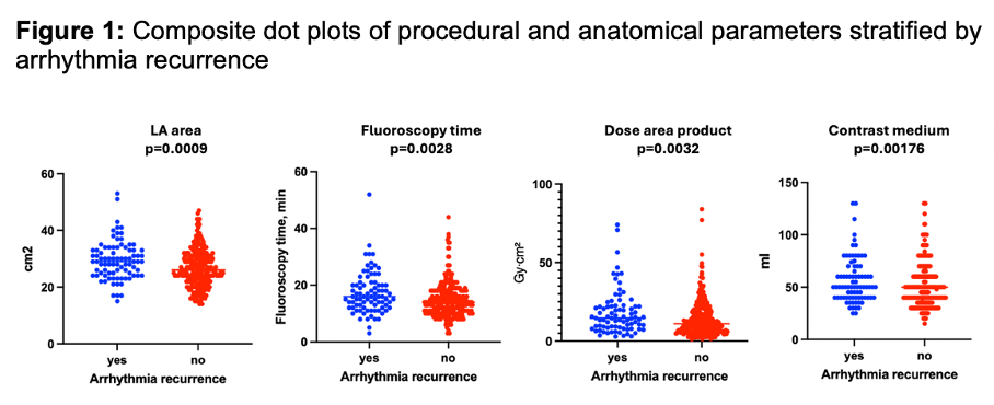
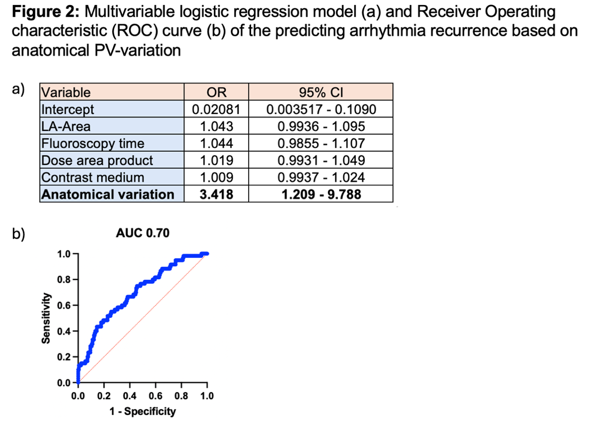
Results: A total of 416 patients (paroxysmal AF: 52.0%, persistent AF: 48%) were followed for 211±174 days. Arrhythmia recurrence occurred in 24.5% of patients. Overall complications occurred in 42 cases (35 minor, 7 major). Univariate analysis for recurrence revealed that among all aforementioned parameters only LA area (p=0.0009), fluoroscopy time (p=0.0028), DAP (p=0.0032), CM volume (p=0.0176), and anatomical variation (p=0.0029) were significant predictors (Fig1). In the multivariable model for recurrence, anatomical variation emerged as the sole independent predictor (OR 3.42, 95% CI 1.21–9.79, p<0.0001, AUC 0.70) for arrhythmia recurrence (Fig2). While DAP was excluded due to high collinearity with fluoroscopy time, the remaining univariately significant parameters were integrated into the score. Fluoroscopy time was also the sole univariate predictor for overall complications (p=0.0303).
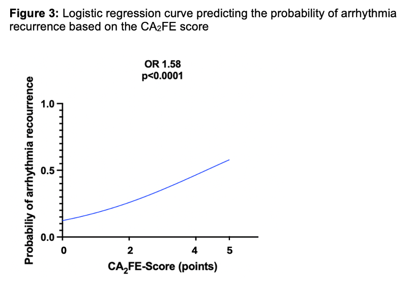
The resulting CA2FE score -weighted as Contrast medium ≥48ml (1 point), Anatomical variation (2 points), Fluoroscopy time ≥14min (1 point), Enlarged LA area ≥27cm2 (1 point)- demonstrated a strong linear prediction of recurrence:
- Score 0: 14.0%
- Score 1: 17.3%
- Score 2: 26.0%
- Score 3: 32.1%
- Score 4: 62.5%
- Score 5: 62.5%
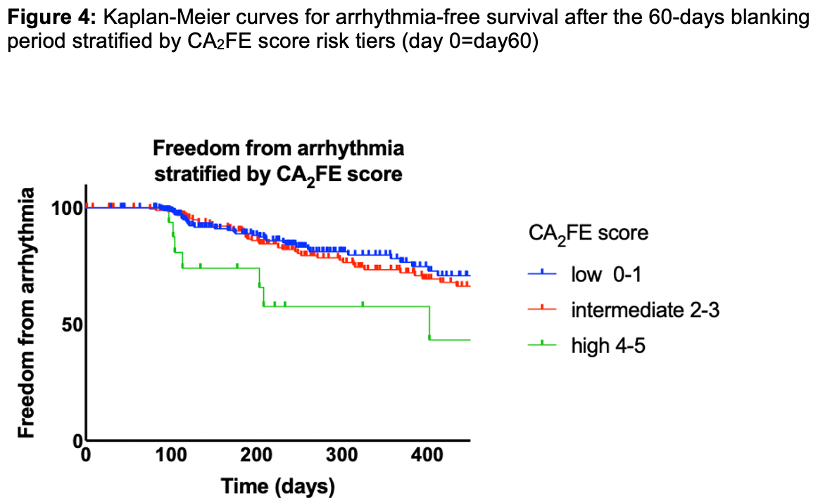
Risk tier stratification (low: 0–1, intermediate: 2–3, high: 4–5) showed highly significant separation in arrhythmia-free survival (p<0.0001; low vs. intermediate: OR 2.01, p=0.0043; low vs. high: OR 8.71, p<0.0001). Each 1-point increase elevated recurrence odds by 58% (OR 1.58, 95% CI 1.30–1.93, p<0.0001; AUC 0.63) (Fig.3 and 4). The score did not predict peri-procedural complications (p=0.20).
Conclusion While PFA provides a highly standardized tool for PVI, procedural and anatomical complexity remains a critical determinant of long-term success. Although limited by an 8.0% lost-to-follow-up rate and changing paradigms like zero-fluoroscopy or contrast-free workflows, this novel 0–5 point score effectively demonstrates that a higher cumulative procedural burden directly correlates with recurrence. It provides an easily calculable clinical tool to stratify post-procedural risk and highlights the need for tailored strategies in complex cases.