Background
The awareness of cardiac amyloidosis (CA) is growing and a steady rise in the number of newly diagnosed is seen. After the approval of therapeutic options pharmaceutical industry pushed awareness, including television advertisements at prime time. Guidelines and position papers have been expanded and refined. Diagnosing CA has become a structured process.
Based on morphologic features, differentiating CA from hypertrophic obstructive cardiomyopathy (HOCM), or non-obstructive variants respectively, is challenging without incorporation of functional parameters.
Aim
We aimed to develop a screening parameter for CA, based on routinely available echocardiographic parameters. Exceptionally we avoided relying on advanced high-tech parameters (Tissue Doppler, 4D, Strain), to provide a broadly accessible screening tool.
To differentiate isolated left ventricular wall thickening from biventricular wall thickening we combined the already well- established parameter „Relative wall thickness“ (RWT) with the thickness of the free wall of the right ventricle (RVfWd). As the RVfWd is physiologically regarded as being half or one third as thick as the walls of the left ventricle (3-5mm), we introduced mathematical weighting factors and exponential transformations to increase its contribution (Table 1).
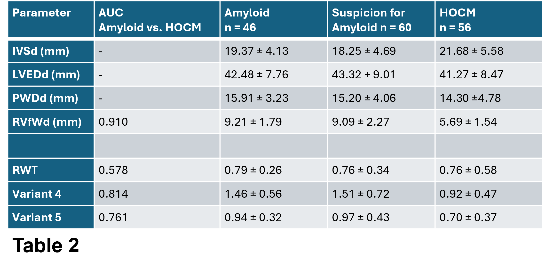
The study cohort comprised pathients with confirmed cardiac Amyloidosis (CA, n=46), suspected cardiac amyloidosis (n=60) and hypertrophic obstructive cardiomyopathy (HOCM, n=56), identified through the Jena Cardiac Hypertrophy Register. In an initial analysis, no significant differences were observed between patients with proven CA and suspected CA. Therfore, both groups were combined and analyzed as a single “CA” group.
Results
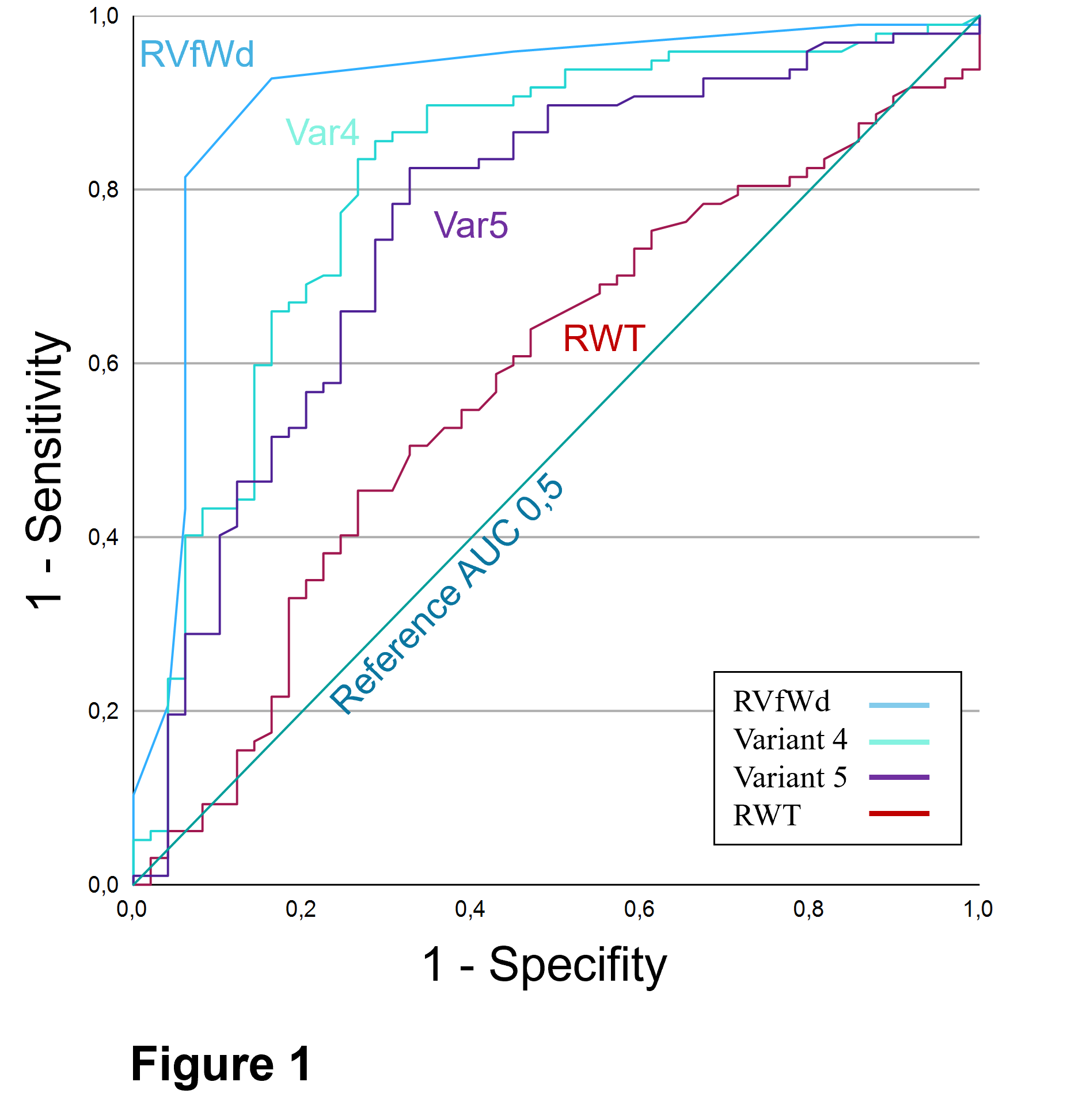
Figure 1 and Table 1 present the ROC curves, AUC and mean values of the investigated parameters. While conventional left ventricular morphological parameters showed comparable values between groups, RVfWd was significantly thinner in HOCM group. Notably, the mean RVfWd in the HOCM group remained above the upper range reported for healthy individuals.
Consistent with these findings, RVfWd alone demonstrated the highest discriminatory power. The diagnostic performance decreased, as the investigated ratios are combined with the left ventricular values. The Variants 1 - 3 were indistinct and therefore omitted from further presentation. Among the proposed composite parameters, variant 4 hat the highest AUC, while Variant 5 had the second highest. Both show good discriminatory performance with AUC values exceeding 0.75.
Conclusion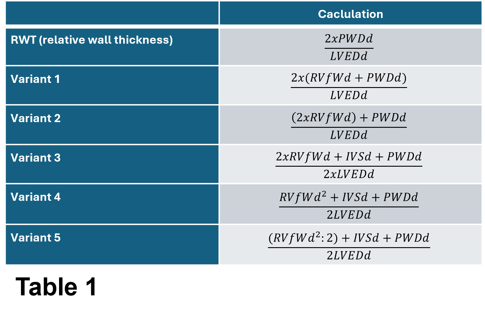
With the objective to go back to simple parameters as screening tools for CA, we developed novel ratios intending to represent biventricular wall thickening. In this proof-of-concept analysis, the proposed parameters were able discriminate between HOCM and CA.
Among the investigated parameters RVfWd alone had the highest discriminatory power. However, its diagnostic performance should be interpreted in the context of the selected study population and requires validation in additional causes of cardiac hypertrophy, which will most likely lead to a downgrading of its power. Further studies will compare the proposed parameters with secondary hypertrophy due to aortic stenosis or arterial hypertension to assess diagnostic value in a broader cilinical and less preselected setting.



