Background:
Myocardial remodeling in aortic valve stenosis (AVS) encompasses left ventricular (LV) hypertrophy, diffuse interstitial fibrosis, replacement fibrosis, and progressive ventricular dysfunction. Although cardiac magnetic resonance imaging (CMR) enables comprehensive assessment of these remodeling processes, its availability remains limited. Circulating biomarkers of myocardial stress, injury, and extracellular matrix remodeling have been associated with adverse outcomes in AVS, but their relationships to specific CMR-defined remodeling phenotypes in AVS remain incompletely understood.
Objectives:
To characterize associations between circulating biomarkers and CMR-derived myocardial remodeling phenotypes in patients with moderate AVS and to explore their relationship with disease progression.
Methods:
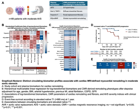
Eighty-nine patients with moderate AVS underwent comprehensive CMR including assessment of LV hypertrophy, native T1, indexed extracellular volume fraction (iECV), and late gadolinium enhancement (LGE). Circulating concentrations of NT-proBNP, high-sensitivity Troponin T, galectin-3 (Gal-3), soluble suppression of tumorigenicity-2 (sST2), and tissue inhibitor of metalloproteinase-1 (TIMP-1) were measured. Associations between biomarkers, CMR phenotypes, echocardiographic AVS severity, and clinical outcomes were analyzed.
Results:
During follow-up, 19 patients (21%) reached the composite endpoint of aortic valve replacement, death, or AS progression. Elevated native T1 (>985 ms) was the only CMR parameter associated with disease progression (OR 3.35; p<0.05) and reduced event-free survival (p=0.049). Native T1 was additionally associated with higher Gal-3 and TIMP-1 concentrations (both p<0.05). Distinct biomarker signatures corresponded to specific remodeling phenotypes. NT-proBNP demonstrated the broadest associations, independently correlating with LV hypertrophy, diffuse fibrosis (native T1 and iECV), replacement fibrosis, and lower LV ejection fraction. Troponin T was independently associated with LGE burden and reduced LV ejection fraction. In contrast, Gal-3 and TIMP-1 showed selective associations with native T1-derived diffuse fibrosis, whereas sST2 demonstrated inverse associations with LV hypertrophy metrics. Hemodynamic AVS severity correlated with adverse CMR remodeling but showed no significant association with circulating biomarker concentrations.
Conclusions:
In moderate AVS, myocardial remodeling is heterogeneous and incompletely captured by valve hemodynamics. Circulating biomarkers map to distinct CMR-defined remodeling phenotypes, with NT-proBNP reflecting global myocardial remodeling, Gal-3 and TIMP-1 identifying diffuse interstitial remodeling, and Troponin T reflecting replacement fibrosis and ventricular dysfunction. Elevated native T1 emerged as the only imaging marker associated with disease progression, supporting diffuse myocardial fibrosis as a key feature of maladaptive remodeling in moderate AVS.